9DW9

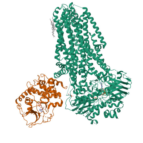
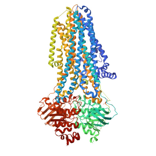
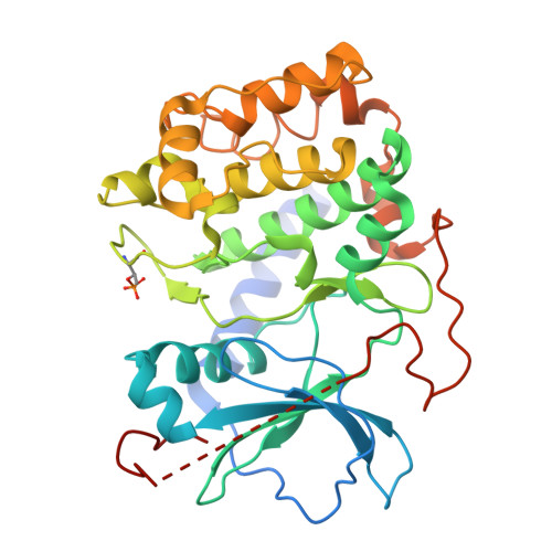
Phosphorylated (E1371Q)CFTR in complex with PKA-C


Experimental Data Snapshot

  • Method: ELECTRON MICROSCOPY
  • Resolution: 2.80 Å
  • Aggregation State: PARTICLE 
  • Reconstruction Method: SINGLE PARTICLE 

wwPDB Validation   3D Report Full Report


This is version 1.2 of the entry. See complete history


Literature

The structures of protein kinase A in complex with CFTR: Mechanisms of phosphorylation and noncatalytic activation.

Fiedorczuk, K.Iordanov, I.Mihalyi, C.Szollosi, A.Csanady, L.Chen, J.

(2024) Proc Natl Acad Sci U S A 121: e2409049121-e2409049121

  • DOI: https://doi.org/10.1073/pnas.2409049121
  • Primary Citation of Related Structures:  
    9DW4, 9DW5, 9DW7, 9DW8, 9DW9

  • PubMed Abstract: 

    Protein kinase A (PKA) is a key regulator of cellular functions by selectively phosphorylating numerous substrates, including ion channels, enzymes, and transcription factors. It has long served as a model system for understanding the eukaryotic kinases. Using cryoelectron microscopy, we present complex structures of the PKA catalytic subunit (PKA-C) bound to a full-length protein substrate, the cystic fibrosis transmembrane conductance regulator (CFTR)-an ion channel vital to human health. CFTR gating requires phosphorylation of its regulatory (R) domain. Unphosphorylated CFTR engages PKA-C at two locations, establishing two "catalytic stations" near to, but not directly involving, the R domain. This configuration, coupled with the conformational flexibility of the R domain, permits transient interactions of the eleven spatially separated phosphorylation sites. Furthermore, we determined two structures of the open-pore CFTR stabilized by PKA-C, providing a molecular basis to understand how PKA-C stimulates CFTR currents even in the absence of phosphorylation.


  • Organizational Affiliation

    Laboratory of Membrane Biology and Biophysics, The Rockefeller University, New York, NY 10065.


Macromolecules
Find similar proteins by:  (by identity cutoff)  |  3D Structure
Entity ID: 1
MoleculeChains Sequence LengthOrganismDetailsImage
Cystic fibrosis transmembrane conductance regulator1,480Homo sapiensMutation(s): 1 
Gene Names: CFTRABCC7
EC: 5.6.1.6
Membrane Entity: Yes 
UniProt & NIH Common Fund Data Resources
Find proteins for P13569 (Homo sapiens)
Explore P13569 
Go to UniProtKB:  P13569
PHAROS:  P13569
GTEx:  ENSG00000001626 
Entity Groups  
Sequence Clusters30% Identity50% Identity70% Identity90% Identity95% Identity100% Identity
UniProt GroupP13569
Sequence Annotations
Expand
  • Reference Sequence
Find similar proteins by:  (by identity cutoff)  |  3D Structure
Entity ID: 2
MoleculeChains Sequence LengthOrganismDetailsImage
cAMP-dependent protein kinase catalytic subunit alphaB [auth G]351Bos taurusMutation(s): 0 
Gene Names: PRKACA
EC: 2.7.11.11
Membrane Entity: Yes 
UniProt
Find proteins for P00517 (Bos taurus)
Explore P00517 
Go to UniProtKB:  P00517
Entity Groups  
Sequence Clusters30% Identity50% Identity70% Identity90% Identity95% Identity100% Identity
UniProt GroupP00517
Sequence Annotations
Expand
  • Reference Sequence
Small Molecules
Ligands 6 Unique
IDChains Name / Formula / InChI Key2D Diagram3D Interactions
ATP (Subject of Investigation/LOI)
Query on ATP

Download Ideal Coordinates CCD File 
G [auth A],
H [auth A],
S [auth G]
ADENOSINE-5'-TRIPHOSPHATE
C10 H16 N5 O13 P3
ZKHQWZAMYRWXGA-KQYNXXCUSA-N
CLR
Query on CLR

Download Ideal Coordinates CCD File 
C [auth A]CHOLESTEROL
C27 H46 O
HVYWMOMLDIMFJA-DPAQBDIFSA-N
UND
Query on UND

Download Ideal Coordinates CCD File 
M [auth A],
N [auth A],
O [auth A]
UNDECANE
C11 H24
RSJKGSCJYJTIGS-UHFFFAOYSA-N
D10
Query on D10

Download Ideal Coordinates CCD File 
D [auth A],
P [auth A],
Q [auth A],
R [auth A]
DECANE
C10 H22
DIOQZVSQGTUSAI-UHFFFAOYSA-N
CL
Query on CL

Download Ideal Coordinates CCD File 
I [auth A],
J [auth A],
K [auth A],
L [auth A]
CHLORIDE ION
Cl
VEXZGXHMUGYJMC-UHFFFAOYSA-M
MG
Query on MG

Download Ideal Coordinates CCD File 
E [auth A],
F [auth A],
T [auth G],
U [auth G]
MAGNESIUM ION
Mg
JLVVSXFLKOJNIY-UHFFFAOYSA-N
Modified Residues  2 Unique
IDChains TypeFormula2D DiagramParent
SEP
Query on SEP
A
L-PEPTIDE LINKINGC3 H8 N O6 PSER
TPO
Query on TPO
B [auth G]L-PEPTIDE LINKINGC4 H10 N O6 PTHR
Experimental Data & Validation

Experimental Data

  • Method: ELECTRON MICROSCOPY
  • Resolution: 2.80 Å
  • Aggregation State: PARTICLE 
  • Reconstruction Method: SINGLE PARTICLE 

Structure Validation

View Full Validation Report


Entry History & Funding Information

Deposition Data


Funding OrganizationLocationGrant Number
Howard Hughes Medical Institute (HHMI)United States--
Cystic Fibrosis FoundationUnited States--

Revision History  (Full details and data files)

  • Version 1.0: 2024-11-13
    Type: Initial release
  • Version 1.1: 2024-11-20
    Changes: Data collection, Database references
  • Version 1.2: 2024-11-27
    Changes: Data collection