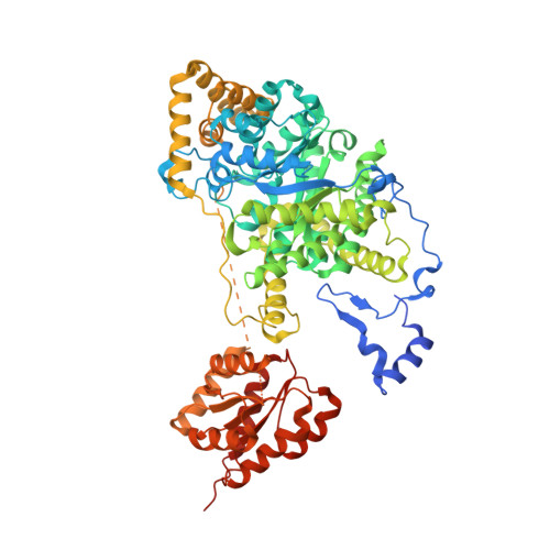
RCSB PDB - 8GJU: Crystal structure of human methylmalonyl-CoA mutase (MMUT) in complex with methylmalonic acidemia type A protein (MMAA), coenzyme A, and GDP

 8GJU

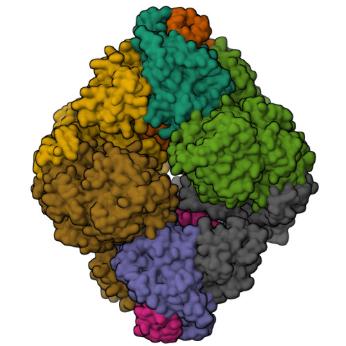
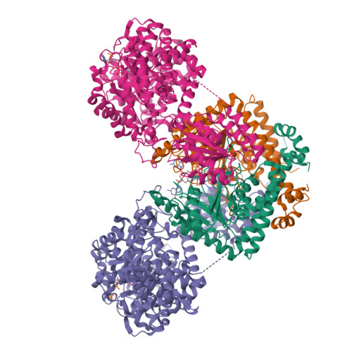
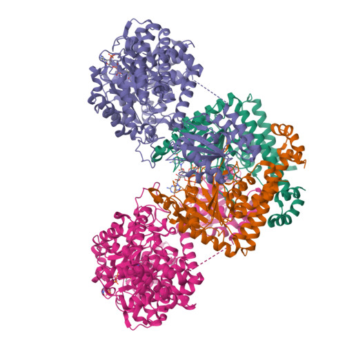
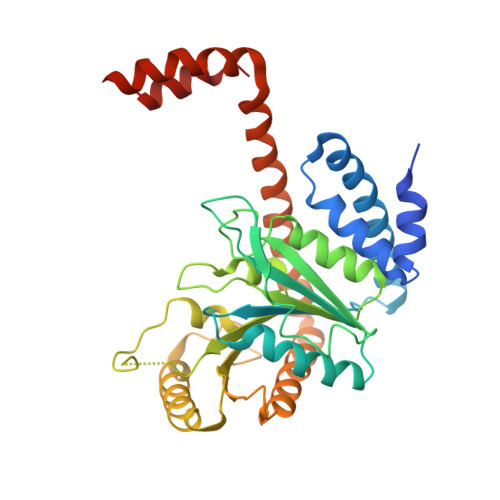
Crystal structure of human methylmalonyl-CoA mutase (MMUT) in complex with methylmalonic acidemia type A protein (MMAA), coenzyme A, and GDP


Experimental Data Snapshot

  • Method: X-RAY DIFFRACTION
  • Resolution: 2.79 Å
  • R-Value Free: 0.276 
  • R-Value Work: 0.229 
  • R-Value Observed: 0.232 

Starting Models: experimental
View more details

wwPDB Validation   3D Report Full Report


Ligand Structure Quality Assessment 

Created with Raphaël 2.3.0Worse 01 BetterLigand structure goodness of fit to experimental dataBest fitted COAClick on this verticalbar to view detailsBest fitted GDPClick on this verticalbar to view details

This is version 1.1 of the entry. See complete history


Literature

Architecture of the human G-protein-methylmalonyl-CoA mutase nanoassembly for B 12 delivery and repair.

Mascarenhas, R.Ruetz, M.Gouda, H.Heitman, N.Yaw, M.Banerjee, R.

(2023) Nat Commun 14: 4332-4332

  • DOI: https://doi.org/10.1038/s41467-023-40077-4
  • Primary Citation of Related Structures:  
    8GJU

  • PubMed Abstract: 

    G-proteins function as molecular switches to power cofactor translocation and confer fidelity in metal trafficking. The G-protein, MMAA, together with MMAB, an adenosyltransferase, orchestrate cofactor delivery and repair of B 12 -dependent human methylmalonyl-CoA mutase (MMUT). The mechanism by which the complex assembles and moves a >1300 Da cargo, or fails in disease, are poorly understood. Herein, we report the crystal structure of the human MMUT-MMAA nano-assembly, which reveals a dramatic 180° rotation of the B 12 domain, exposing it to solvent. The complex, stabilized by MMAA wedging between two MMUT domains, leads to ordering of the switch I and III loops, revealing the molecular basis of mutase-dependent GTPase activation. The structure explains the biochemical penalties incurred by methylmalonic aciduria-causing mutations that reside at the MMAA-MMUT interfaces we identify here.


  • Organizational Affiliation

    Department of Biological Chemistry, University of Michigan, Ann Arbor, MI, 48109, USA.


Macromolecules
Find similar proteins by:  (by identity cutoff)  |  3D Structure
Entity ID: 1
MoleculeChains Sequence LengthOrganismDetailsImage
Methylmalonic aciduria type A protein, mitochondrialA [auth D],
B [auth F],
F [auth A],
G [auth B]
349Homo sapiensMutation(s): 0 
Gene Names: MMAA
EC: 3.6
UniProt & NIH Common Fund Data Resources
Find proteins for Q8IVH4 (Homo sapiens)
Explore Q8IVH4 
Go to UniProtKB:  Q8IVH4
PHAROS:  Q8IVH4
GTEx:  ENSG00000151611 
Entity Groups  
Sequence Clusters30% Identity50% Identity70% Identity90% Identity95% Identity100% Identity
UniProt GroupQ8IVH4
Sequence Annotations
Expand
  • Reference Sequence
Find similar proteins by:  (by identity cutoff)  |  3D Structure
Entity ID: 2
MoleculeChains Sequence LengthOrganismDetailsImage
Methylmalonyl-CoA mutase, mitochondrialC [auth J],
D [auth K],
E [auth L],
H
748Homo sapiensMutation(s): 0 
Gene Names: MUT
EC: 5.4.99.2
UniProt & NIH Common Fund Data Resources
Find proteins for P22033 (Homo sapiens)
Explore P22033 
Go to UniProtKB:  P22033
PHAROS:  P22033
GTEx:  ENSG00000146085 
Entity Groups  
Sequence Clusters30% Identity50% Identity70% Identity90% Identity95% Identity100% Identity
UniProt GroupP22033
Sequence Annotations
Expand
  • Reference Sequence
Small Molecules
Ligands 3 Unique
IDChains Name / Formula / InChI Key2D Diagram3D Interactions
COA (Subject of Investigation/LOI)
Query on COA

Download Ideal Coordinates CCD File 
M [auth J],
N [auth K],
O [auth L],
T [auth H]
COENZYME A
C21 H36 N7 O16 P3 S
RGJOEKWQDUBAIZ-IBOSZNHHSA-N
GDP (Subject of Investigation/LOI)
Query on GDP

Download Ideal Coordinates CCD File 
I [auth D],
K [auth F],
P [auth A],
R [auth B]
GUANOSINE-5'-DIPHOSPHATE
C10 H15 N5 O11 P2
QGWNDRXFNXRZMB-UUOKFMHZSA-N
MG
Query on MG

Download Ideal Coordinates CCD File 
J [auth D],
L [auth F],
Q [auth A],
S [auth B]
MAGNESIUM ION
Mg
JLVVSXFLKOJNIY-UHFFFAOYSA-N
Experimental Data & Validation

Experimental Data

  • Method: X-RAY DIFFRACTION
  • Resolution: 2.79 Å
  • R-Value Free: 0.276 
  • R-Value Work: 0.229 
  • R-Value Observed: 0.232 
  • Space Group: P 1 21 1
Unit Cell:
Length ( Å )Angle ( ˚ )
a = 95.806α = 90
b = 221.872β = 105.53
c = 121.824γ = 90
Software Package:
Software NamePurpose
PHENIXrefinement
XDSdata reduction
XDSdata scaling
PHASERphasing

Structure Validation

View Full Validation Report


Ligand Structure Quality Assessment 

Created with Raphaël 2.3.0Worse 01 BetterLigand structure goodness of fit to experimental dataBest fitted COAClick on this verticalbar to view detailsBest fitted GDPClick on this verticalbar to view details

Entry History & Funding Information

Deposition Data


Funding OrganizationLocationGrant Number
National Institutes of Health/National Institute of Diabetes and Digestive and Kidney Disease (NIH/NIDDK)United StatesDK45776
National Institutes of Health/National Institute of General Medical Sciences (NIH/NIGMS)United StatesK99-GM1434820

Revision History  (Full details and data files)

  • Version 1.0: 2023-08-09
    Type: Initial release
  • Version 1.1: 2024-05-22
    Changes: Data collection