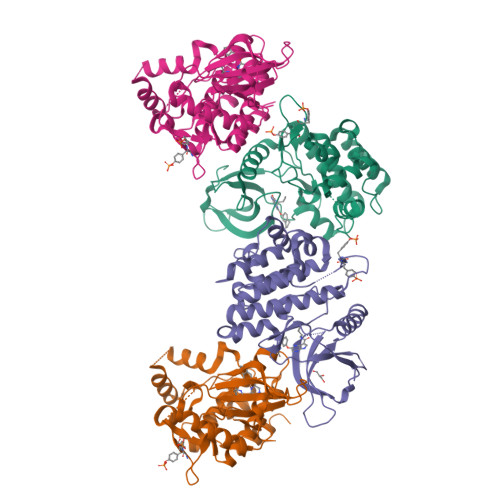
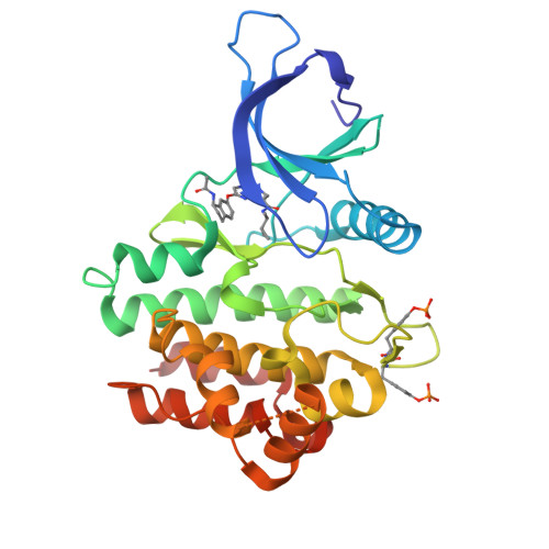
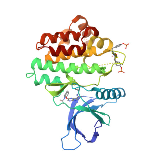
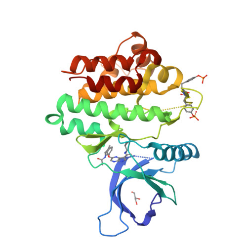
3ZEP | pdb_00003zep

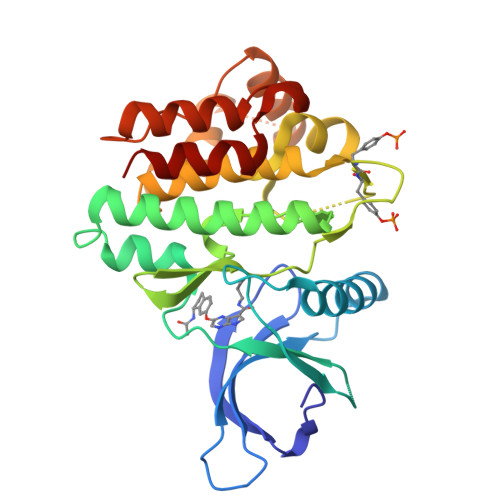
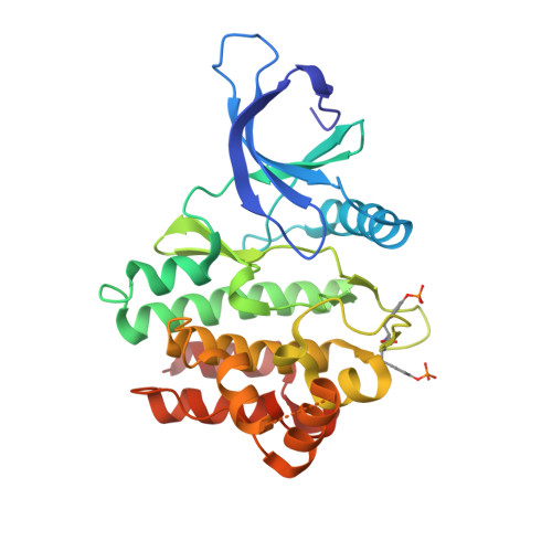
Crystal Structure of JAK3 Kinase Domain in Complex with a Pyrrolopyrazine-2-phenyl Ether Inhibitor


Experimental Data Snapshot

  • Method: X-RAY DIFFRACTION
  • Resolution: 2.35 Å
  • R-Value Free: 
    0.265 (Depositor), 0.268 (DCC) 
  • R-Value Work: 
    0.226 (Depositor), 0.227 (DCC) 
  • R-Value Observed: 
    0.226 (Depositor) 

wwPDB Validation   3D Report Full Report


Ligand Structure Quality Assessment 

Created with Raphaël 2.3.0Worse 01 BetterLigand structure goodness of fit to experimental dataBest fitted 1NXClick on this verticalbar to view details

This is version 1.2 of the entry. See complete history


Literature

Discovery of a Series of Novel 5H-Pyrrolo[2,3-B]Pyrazine-2-Phenyl Ethers, as Potent Jak3 Kinase Inhibitors.

Jaime-Figueroa, S.De Vicente, J.Hermann, J.Jahangir, A.Jin, S.Kuglstatter, A.Lynch, S.M.Menke, J.Niu, L.Patel, V.Shao, A.Soth, M.Vu, M.D.Yee, C.

(2013) Bioorg Med Chem Lett 23: 2522

  • DOI: https://doi.org/10.1016/j.bmcl.2013.03.015
  • Primary Citation of Related Structures:  
    3ZEP, 4I6Q

  • PubMed Abstract: 

    We report the discovery of a novel series of ATP-competitive Janus kinase 3 (JAK3) inhibitors based on the 5H-pyrrolo[2,3-b]pyrazine scaffold. The initial leads in this series, compounds 1a and 1h, showed promising potencies, but a lack of selectivity against other isoforms in the JAK family. Computational and crystallographic analysis suggested that the phenyl ether moiety possessed a favorable vector to achieve selectivity. Exploration of this vector resulted in the identification of 12b and 12d, as potent JAK3 inhibitors, demonstrating improved JAK family and kinase selectivity.


  • Organizational Affiliation

    Hoffmann-La Roche Inc., pRED, Pharma Research & Early Development, Small Molecule Research, Discovery Chemistry, 340 Kingsland Street, Nutley, NJ 07110-1199, USA. saul.jaime@roche.com


Macromolecules
Find similar proteins by:  (by identity cutoff)  |  3D Structure
Entity ID: 1
MoleculeChains Sequence LengthOrganismDetailsImage
TYROSINE-PROTEIN KINASE JAK3
A, B, C, D
288Homo sapiensMutation(s): 2 
EC: 2.7.10.2 (PDB Primary Data), 2.7.1.112 (PDB Primary Data)
UniProt & NIH Common Fund Data Resources
Find proteins for P52333 (Homo sapiens)
Explore P52333 
Go to UniProtKB:  P52333
PHAROS:  P52333
GTEx:  ENSG00000105639 
Entity Groups  
Sequence Clusters30% Identity50% Identity70% Identity90% Identity95% Identity100% Identity
UniProt GroupP52333
Sequence Annotations
Expand
  • Reference Sequence
Small Molecules
Modified Residues  1 Unique
IDChains TypeFormula2D DiagramParent
PTR
Query on PTR
A, B, C, D
L-PEPTIDE LINKINGC9 H12 N O6 PTYR
Binding Affinity Annotations 
IDSourceBinding Affinity
1NX BindingDB:  3ZEP IC50: min: 22, max: 700 (nM) from 2 assay(s)
Experimental Data & Validation

Experimental Data

  • Method: X-RAY DIFFRACTION
  • Resolution: 2.35 Å
  • R-Value Free:  0.265 (Depositor), 0.268 (DCC) 
  • R-Value Work:  0.226 (Depositor), 0.227 (DCC) 
  • R-Value Observed: 0.226 (Depositor) 
Space Group: P 1 21 1
Unit Cell:
Length ( Å )Angle ( ˚ )
a = 57.554α = 90
b = 114.076β = 96.82
c = 104.196γ = 90
Software Package:
Software NamePurpose
REFMACrefinement
XDSdata reduction
SCALAdata scaling
REFMACphasing

Structure Validation

View Full Validation Report


Ligand Structure Quality Assessment 

Created with Raphaël 2.3.0Worse 01 BetterLigand structure goodness of fit to experimental dataBest fitted 1NXClick on this verticalbar to view details

Entry History 

Deposition Data

Revision History  (Full details and data files)

  • Version 1.0: 2013-12-11
    Type: Initial release
  • Version 1.1: 2016-09-14
    Changes: Database references
  • Version 1.2: 2024-10-23
    Changes: Data collection, Database references, Derived calculations, Other, Structure summary