3LDQ

Crystal structure of HSC70/BAG1 in complex with small molecule inhibitor


Experimental Data Snapshot

  • Method: X-RAY DIFFRACTION
  • Resolution: 1.90 Å
  • R-Value Free: 
    0.256 (Depositor), 0.250 (DCC) 
  • R-Value Work: 
    0.203 (Depositor), 0.200 (DCC) 
  • R-Value Observed: 
    0.206 (Depositor) 

Starting Model: experimental
View more details

wwPDB Validation   3D Report Full Report


Ligand Structure Quality Assessment 

Created with Raphaël 2.3.0Worse 01 BetterLigand structure goodness of fit to experimental dataBest fitted 3P1Click on this verticalbar to view details

This is version 1.3 of the entry. See complete history


Literature

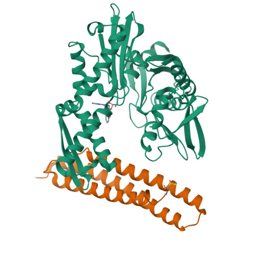
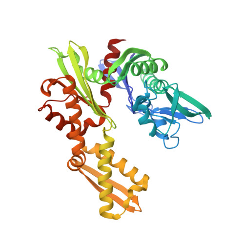
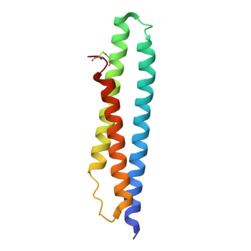
Adenosine-Derived Inhibitors of 78 kDa Glucose Regulated Protein (Grp78) ATPase: Insights into Isoform Selectivity.

Macias, A.T.Williamson, D.S.Allen, N.Borgognoni, J.Clay, A.Daniels, Z.Dokurno, P.Drysdale, M.J.Francis, G.L.Graham, C.J.Howes, R.Matassova, N.Murray, J.B.Parsons, R.Shaw, T.Surgenor, A.E.Terry, L.Wang, Y.Wood, M.Massey, A.J.

(2011) J Med Chem 54: 4034-4041

  • DOI: https://doi.org/10.1021/jm101625x
  • Primary Citation of Related Structures:  
    3LDL, 3LDN, 3LDO, 3LDP, 3LDQ, 3M3Z

  • PubMed Abstract: 

    78 kDa glucose-regulated protein (Grp78) is a heat shock protein (HSP) involved in protein folding that plays a role in cancer cell proliferation. Binding of adenosine-derived inhibitors to Grp78 was characterized by surface plasmon resonance and isothermal titration calorimetry. The most potent compounds were 13 (VER-155008) with K(D) = 80 nM and 14 with K(D) = 60 nM. X-ray crystal structures of Grp78 bound to ATP, ADPnP, and adenosine derivative 10 revealed differences in the binding site between Grp78 and homologous proteins.


  • Organizational Affiliation

    Vernalis (R&D) Ltd., Granta Park, Great Abington, Cambridge, CB21 6GB, UK. a.macias@vernalis.com


Macromolecules
Find similar proteins by:  (by identity cutoff)  |  3D Structure
Entity ID: 1
MoleculeChains Sequence LengthOrganismDetailsImage
Heat shock cognate 71 kDa protein381Homo sapiensMutation(s): 0 
Gene Names: HSPA8HSC70HSP73HSPA10
EC: 3.6.4.10
UniProt & NIH Common Fund Data Resources
Find proteins for P11142 (Homo sapiens)
Explore P11142 
Go to UniProtKB:  P11142
PHAROS:  P11142
GTEx:  ENSG00000109971 
Entity Groups  
Sequence Clusters30% Identity50% Identity70% Identity90% Identity95% Identity100% Identity
UniProt GroupP11142
Sequence Annotations
Expand
  • Reference Sequence
Find similar proteins by:  (by identity cutoff)  |  3D Structure
Entity ID: 2
MoleculeChains Sequence LengthOrganismDetailsImage
BAG family molecular chaperone regulator 1114Homo sapiensMutation(s): 0 
Gene Names: BAG1HAP
UniProt & NIH Common Fund Data Resources
Find proteins for Q99933 (Homo sapiens)
Explore Q99933 
Go to UniProtKB:  Q99933
PHAROS:  Q99933
GTEx:  ENSG00000107262 
Entity Groups  
Sequence Clusters30% Identity50% Identity70% Identity90% Identity95% Identity100% Identity
UniProt GroupQ99933
Sequence Annotations
Expand
  • Reference Sequence
Small Molecules
Ligands 1 Unique
IDChains Name / Formula / InChI Key2D Diagram3D Interactions
3P1
Query on 3P1

Download Ideal Coordinates CCD File 
C [auth A]8-[(quinolin-2-ylmethyl)amino]adenosine
C20 H21 N7 O4
NAHSCHKAPXMNFP-NVQRDWNXSA-N
Binding Affinity Annotations 
IDSourceBinding Affinity
3P1 BindingDB:  3LDQ Ki: 4320 (nM) from 1 assay(s)
Experimental Data & Validation

Experimental Data

  • Method: X-RAY DIFFRACTION
  • Resolution: 1.90 Å
  • R-Value Free:  0.256 (Depositor), 0.250 (DCC) 
  • R-Value Work:  0.203 (Depositor), 0.200 (DCC) 
  • R-Value Observed: 0.206 (Depositor) 
Space Group: P 1 21 1
Unit Cell:
Length ( Å )Angle ( ˚ )
a = 40.774α = 90
b = 121.995β = 106.88
c = 54.25γ = 90
Software Package:
Software NamePurpose
MxCuBEdata collection
MOLREPphasing
REFMACrefinement
HKL-2000data reduction
HKL-2000data scaling

Structure Validation

View Full Validation Report


Ligand Structure Quality Assessment 

Created with Raphaël 2.3.0Worse 01 BetterLigand structure goodness of fit to experimental dataBest fitted 3P1Click on this verticalbar to view details

Entry History 

Deposition Data

Revision History  (Full details and data files)

  • Version 1.0: 2011-01-26
    Type: Initial release
  • Version 1.1: 2011-07-13
    Changes: Version format compliance
  • Version 1.2: 2023-09-06
    Changes: Data collection, Database references, Derived calculations, Refinement description
  • Version 1.3: 2024-10-09
    Changes: Structure summary