2PE1 | pdb_00002pe1

CRYSTAL STRUCTURE OF HUMAN PHOSPHOINOSITIDE-DEPENDENT PROTEIN KINASE 1 (PDK1) {2-Oxo-3-[1-(1H-pyrrol-2-yl)-eth-(Z)-ylidene]-2,3-dihydro-1H-indol-5-yl}-urea {BX-517} COMPLEX


Experimental Data Snapshot

  • Method: X-RAY DIFFRACTION
  • Resolution: 2.14 Å
  • R-Value Free: 
    0.282 (Depositor), 0.287 (DCC) 
  • R-Value Work: 
    0.222 (Depositor), 0.224 (DCC) 
  • R-Value Observed: 
    0.224 (Depositor) 

wwPDB Validation   3D Report Full Report


Ligand Structure Quality Assessment 

Created with Raphaël 2.3.0Worse 01 BetterLigand structure goodness of fit to experimental dataBest fitted 517Click on this verticalbar to view details

This is version 1.3 of the entry. See complete history


Literature

Indolinone based phosphoinositide-dependent kinase-1 (PDK1) inhibitors. Part 1: Design, synthesis and biological activity.

Islam, I.Bryant, J.Chou, Y.L.Kochanny, M.J.Lee, W.Phillips, G.B.Yu, H.Adler, M.Whitlow, M.Ho, E.Lentz, D.Polokoff, M.A.Subramanyam, B.Wu, J.M.Zhu, D.Feldman, R.I.Arnaiz, D.O.

(2007) Bioorg Med Chem Lett 17: 3814-3818

  • DOI: https://doi.org/10.1016/j.bmcl.2007.04.071
  • Primary Citation of Related Structures:  
    2PE0, 2PE1

  • PubMed Abstract: 

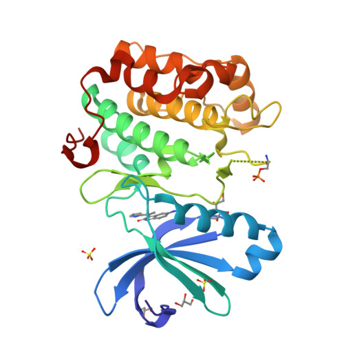
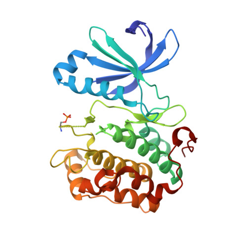
    HTS screening identified 1 with micromolar inhibitory activity against PDK1. Optimization of 1 afforded 4i (BX-517) which has single-digit nanomolar activity against PDK1 and excellent selectivity against PKA.


  • Organizational Affiliation

    Berlex Biosciences, 2600 Hilltop Dr. Richmond, CA 94804, USA.


Macromolecules
Find similar proteins by:  (by identity cutoff)  |  3D Structure
Entity ID: 1
MoleculeChains Sequence LengthOrganismDetailsImage
3-phosphoinositide-dependent protein kinase 1286Homo sapiensMutation(s): 1 
Gene Names: PDPK1PDK1
EC: 2.7.11.1
UniProt & NIH Common Fund Data Resources
Find proteins for O15530 (Homo sapiens)
Explore O15530 
Go to UniProtKB:  O15530
PHAROS:  O15530
GTEx:  ENSG00000140992 
Entity Groups  
Sequence Clusters30% Identity50% Identity70% Identity90% Identity95% Identity100% Identity
UniProt GroupO15530
Sequence Annotations
Expand
  • Reference Sequence
Small Molecules
Ligands 3 Unique
IDChains Name / Formula / InChI Key2D Diagram3D Interactions
517
Query on 517

Download Ideal Coordinates CCD File 
D [auth A]1-{2-OXO-3-[(1R)-1-(1H-PYRROL-2-YL)ETHYL]-2H-INDOL-5-YL}UREA
C15 H14 N4 O2
KJDBLWKTHMHALD-QMMMGPOBSA-N
SO4
Query on SO4

Download Ideal Coordinates CCD File 
B [auth A],
C [auth A]
SULFATE ION
O4 S
QAOWNCQODCNURD-UHFFFAOYSA-L
GOL
Query on GOL

Download Ideal Coordinates CCD File 
E [auth A],
F [auth A],
G [auth A],
H [auth A]
GLYCEROL
C3 H8 O3
PEDCQBHIVMGVHV-UHFFFAOYSA-N
Modified Residues  1 Unique
IDChains TypeFormula2D DiagramParent
SEP
Query on SEP
A
L-PEPTIDE LINKINGC3 H8 N O6 PSER
Experimental Data & Validation

Experimental Data

  • Method: X-RAY DIFFRACTION
  • Resolution: 2.14 Å
  • R-Value Free:  0.282 (Depositor), 0.287 (DCC) 
  • R-Value Work:  0.222 (Depositor), 0.224 (DCC) 
  • R-Value Observed: 0.224 (Depositor) 
Space Group: P 32 2 1
Unit Cell:
Length ( Å )Angle ( ˚ )
a = 125.234α = 90
b = 125.234β = 90
c = 47.696γ = 120
Software Package:
Software NamePurpose
CNXrefinement
XTALVIEWrefinement
X-GENdata reduction
X-GENdata scaling
CNXphasing

Structure Validation

View Full Validation Report


Ligand Structure Quality Assessment 

Created with Raphaël 2.3.0Worse 01 BetterLigand structure goodness of fit to experimental dataBest fitted 517Click on this verticalbar to view details

Entry History 

Deposition Data

Revision History  (Full details and data files)

  • Version 1.0: 2007-06-12
    Type: Initial release
  • Version 1.1: 2008-05-01
    Changes: Version format compliance
  • Version 1.2: 2011-07-13
    Changes: Non-polymer description, Version format compliance
  • Version 1.3: 2024-11-06
    Changes: Data collection, Database references, Derived calculations, Structure summary