9FZ2 | pdb_00009fz2

Crystal structure of Amylase 5 (Amy5) from Ruminococcus bromii covalently bound to alpha-1,6 branched pseudo-trisaccharide activity-based probe


Experimental Data Snapshot

  • Method: X-RAY DIFFRACTION
  • Resolution: 1.40 Å
  • R-Value Free: 
    0.222 (Depositor), 0.231 (DCC) 
  • R-Value Work: 
    0.187 (Depositor), 0.196 (DCC) 
  • R-Value Observed: 
    0.189 (Depositor) 

Starting Model: experimental
View more details

wwPDB Validation   3D Report Full Report


Ligand Structure Quality Assessment 

Created with Raphaël 2.3.0Worse 01 BetterLigand structure goodness of fit to experimental dataBest fitted PBWClick on this verticalbar to view detailsBest fitted A1IHIClick on this verticalbar to view details

This is version 1.1 of the entry. See complete history


Literature

Precision Activity-Based alpha-Amylase Probes for Dissection and Annotation of Linear and Branched-Chain Starch-Degrading Enzymes.

Pickles, I.B.Chen, Y.Moroz, O.Brown, H.A.de Boer, C.Armstrong, Z.McGregor, N.G.S.Artola, M.Codee, J.D.C.Koropatkin, N.M.Overkleeft, H.S.Davies, G.J.

(2025) Angew Chem Int Ed Engl 64: e202415219-e202415219

  • DOI: https://doi.org/10.1002/anie.202415219
  • Primary Citation of Related Structures:  
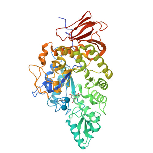
    9FYZ, 9FZ0, 9FZ2, 9FZ3

  • PubMed Abstract: 

    α-Amylases are the workhorse enzymes of starch degradation. They are central to human health, including as targets for anti-diabetic compounds, but are also the key enzymes in the industrial processing of starch for biofuels, corn syrups, brewing and detergents. Dissection of the activity, specificity and stability of α-amylases is crucial to understanding their biology and allowing their exploitation. Yet, functional characterization lags behind DNA sequencing and genomics; and new tools are required for rapid analysis of α-amylase function. Here, we design, synthesize and apply new branched α-amylase activity-based probes. Using both α-1,6 branched and unbranched α-1,4 maltobiose activity-based probes we were able to explore the stability and substrate specificity of both a panel of human gut microbial α-amylases and a panel of industrially relevant α-amylases. We also demonstrate how we can detect and annotate the substrate specificity of α-amylases in the complex cell lysate of both a prominent gut microbe and a diverse compost sample by in-gel fluorescence and mass spectrometry. A toolbox of starch-active activity-based probes will enable rapid functional dissection of α-amylases. We envisage activity-based probes contributing to better selection and engineering of enzymes for industrial application as well as fundamental analysis of enzymes in human health.


  • Organizational Affiliation

    University of York, York Structural Biology Laboratory, Department of Chemistry, UNITED KINGDOM OF GREAT BRITAIN AND NORTHERN IRELAND.


Macromolecules
Find similar proteins by:  (by identity cutoff)  |  3D Structure
Entity ID: 1
MoleculeChains Sequence LengthOrganismDetailsImage
Alpha-amylase542Ruminococcus bromiiMutation(s): 0 
Gene Names: RBL236_00453
EC: 3.2.1.1
UniProt
Find proteins for A0A2N0UJ68 (Ruminococcus bromii)
Explore A0A2N0UJ68 
Go to UniProtKB:  A0A2N0UJ68
Entity Groups  
Sequence Clusters30% Identity50% Identity70% Identity90% Identity95% Identity100% Identity
UniProt GroupA0A2N0UJ68
Sequence Annotations
Expand
  • Reference Sequence
Oligosaccharides

Help

Entity ID: 2
MoleculeChains Length2D Diagram Glycosylation3D Interactions
alpha-D-glucopyranose-(1-6)-alpha-D-glucopyranose
B
2N/A
Glycosylation Resources
GlyTouCan:  G69864PN
GlyCosmos:  G69864PN
Experimental Data & Validation

Experimental Data

  • Method: X-RAY DIFFRACTION
  • Resolution: 1.40 Å
  • R-Value Free:  0.222 (Depositor), 0.231 (DCC) 
  • R-Value Work:  0.187 (Depositor), 0.196 (DCC) 
  • R-Value Observed: 0.189 (Depositor) 
Space Group: P 1 21 1
Unit Cell:
Length ( Å )Angle ( ˚ )
a = 49.869α = 90
b = 94.873β = 94.64
c = 49.904γ = 90
Software Package:
Software NamePurpose
REFMACrefinement
DIALSdata reduction
Aimlessdata scaling
PHASERphasing

Structure Validation

View Full Validation Report


Ligand Structure Quality Assessment 

Created with Raphaël 2.3.0Worse 01 BetterLigand structure goodness of fit to experimental dataBest fitted PBWClick on this verticalbar to view detailsBest fitted A1IHIClick on this verticalbar to view details

Entry History & Funding Information

Deposition Data


Funding OrganizationLocationGrant Number
European Research Council (ERC)European Union951231

Revision History  (Full details and data files)

  • Version 1.0: 2024-12-11
    Type: Initial release
  • Version 1.1: 2025-02-05
    Changes: Database references