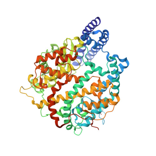
8TOS

ACE2-peptide 6 complex


Experimental Data Snapshot

  • Method: X-RAY DIFFRACTION
  • Resolution: 2.35 Å
  • R-Value Free: 0.200 
  • R-Value Work: 0.182 
  • R-Value Observed: 0.183 

Starting Model: experimental
View more details

wwPDB Validation   3D Report Full Report


Ligand Structure Quality Assessment 


This is version 1.1 of the entry. See complete history


Literature

Discovery of High Affinity Cyclic Peptide Ligands for Human ACE2 with SARS-CoV-2 Entry Inhibitory Activity.

Bedding, M.J.Franck, C.Johansen-Leete, J.Aggarwal, A.Maxwell, J.W.C.Patel, K.Hawkins, P.M.E.Low, J.K.K.Siddiquee, R.Sani, H.M.Ford, D.J.Turville, S.Mackay, J.P.Passioura, T.Christie, M.Payne, R.J.

(2024) ACS Chem Biol 19: 141-152

  • DOI: https://doi.org/10.1021/acschembio.3c00568
  • Primary Citation of Related Structures:  
    8TOQ, 8TOR, 8TOS, 8TOT, 8TOU

  • PubMed Abstract: 

    The development of effective antiviral compounds is essential for mitigating the effects of the COVID-19 pandemic. Entry of SARS-CoV-2 virions into host cells is mediated by the interaction between the viral spike (S) protein and membrane-bound angiotensin-converting enzyme 2 (ACE2) on the surface of epithelial cells. Inhibition of this viral protein-host protein interaction is an attractive avenue for the development of antiviral molecules with numerous spike-binding molecules generated to date. Herein, we describe an alternative approach to inhibit the spike-ACE2 interaction by targeting the spike-binding interface of human ACE2 via mRNA display. Two consecutive display selections were performed to direct cyclic peptide ligand binding toward the spike binding interface of ACE2. Through this process, potent cyclic peptide binders of human ACE2 (with affinities in the picomolar to nanomolar range) were identified, two of which neutralized SARS-CoV-2 entry. This work demonstrates the potential of targeting ACE2 for the generation of anti-SARS-CoV-2 therapeutics as well as broad spectrum antivirals for the treatment of SARS-like betacoronavirus infection.


  • Organizational Affiliation

    School of Chemistry, The University of Sydney, Sydney, New South Wales 2006, Australia.


Macromolecules
Find similar proteins by:  (by identity cutoff)  |  3D Structure
Entity ID: 1
MoleculeChains Sequence LengthOrganismDetailsImage
Angiotensin-converting enzyme 2625Homo sapiensMutation(s): 0 
Gene Names: ACE2
EC: 3.4.17 (UniProt), 3.4.17.23 (UniProt)
UniProt & NIH Common Fund Data Resources
Find proteins for Q9BYF1 (Homo sapiens)
Explore Q9BYF1 
Go to UniProtKB:  Q9BYF1
PHAROS:  Q9BYF1
GTEx:  ENSG00000130234 
Entity Groups  
Sequence Clusters30% Identity50% Identity70% Identity90% Identity95% Identity100% Identity
UniProt GroupQ9BYF1
Glycosylation
Glycosylation Sites: 3Go to GlyGen: Q9BYF1-1
Sequence Annotations
Expand
  • Reference Sequence

Find similar proteins by:  Sequence   |   3D Structure  

Entity ID: 2
MoleculeChains Sequence LengthOrganismDetailsImage
Peptide 6B [auth E]17synthetic constructMutation(s): 0 
Entity Groups  
Sequence Clusters30% Identity50% Identity70% Identity90% Identity95% Identity100% Identity
Sequence Annotations
Expand
  • Reference Sequence
Oligosaccharides

Help

Entity ID: 3
MoleculeChains Length2D Diagram Glycosylation3D Interactions
alpha-L-fucopyranose-(1-6)-2-acetamido-2-deoxy-beta-D-glucopyranoseC [auth B],
D [auth C]
2N-Glycosylation
Glycosylation Resources
GlyTouCan:  G86851RC
GlyCosmos:  G86851RC
GlyGen:  G86851RC
Small Molecules
Ligands 4 Unique
IDChains Name / Formula / InChI Key2D Diagram3D Interactions
NAG
Query on NAG

Download Ideal Coordinates CCD File 
G [auth A]2-acetamido-2-deoxy-beta-D-glucopyranose
C8 H15 N O6
OVRNDRQMDRJTHS-FMDGEEDCSA-N
GOL
Query on GOL

Download Ideal Coordinates CCD File 
E [auth A],
F [auth A],
H [auth A],
I [auth A],
J [auth A]
GLYCEROL
C3 H8 O3
PEDCQBHIVMGVHV-UHFFFAOYSA-N
ZN
Query on ZN

Download Ideal Coordinates CCD File 
K [auth A]ZINC ION
Zn
PTFCDOFLOPIGGS-UHFFFAOYSA-N
CL
Query on CL

Download Ideal Coordinates CCD File 
L [auth A]CHLORIDE ION
Cl
VEXZGXHMUGYJMC-UHFFFAOYSA-M
Experimental Data & Validation

Experimental Data

  • Method: X-RAY DIFFRACTION
  • Resolution: 2.35 Å
  • R-Value Free: 0.200 
  • R-Value Work: 0.182 
  • R-Value Observed: 0.183 
  • Space Group: H 3
Unit Cell:
Length ( Å )Angle ( ˚ )
a = 179.566α = 90
b = 179.566β = 90
c = 70.252γ = 120
Software Package:
Software NamePurpose
Cootmodel building
PHENIXrefinement
XDSdata reduction
Aimlessdata scaling
PHASERphasing

Structure Validation

View Full Validation Report


Ligand Structure Quality Assessment 


Entry History & Funding Information

Deposition Data


Funding OrganizationLocationGrant Number
Australian Research Council (ARC)AustraliaDP220102497
Australian Research Council (ARC)AustraliaCE200100012
National Health and Medical Research Council (NHMRC, Australia)AustraliaAPP1141142
National Health and Medical Research Council (NHMRC, Australia)AustraliaAPP1153493

Revision History  (Full details and data files)

  • Version 1.0: 2024-02-07
    Type: Initial release
  • Version 1.1: 2024-10-23
    Changes: Structure summary