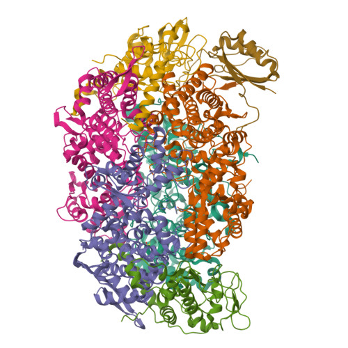
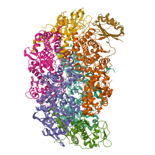
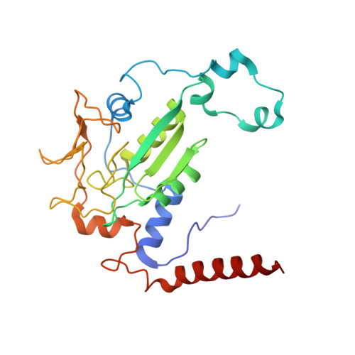
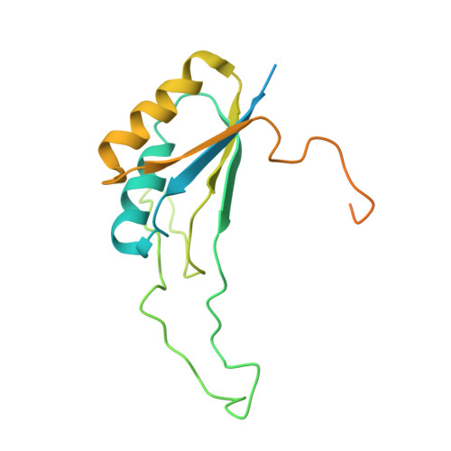
8GF6

Apo-apo MCR assembly intermediate


Experimental Data Snapshot

  • Method: ELECTRON MICROSCOPY
  • Resolution: 3.10 Å
  • Aggregation State: PARTICLE 
  • Reconstruction Method: SINGLE PARTICLE 

wwPDB Validation   3D Report Full Report


This is version 1.0 of the entry. See complete history


Literature

McrD binds asymmetrically to methyl-coenzyme M reductase improving active-site accessibility during assembly.

Chadwick, G.L.Joiner, A.M.N.Ramesh, S.Mitchell, D.A.Nayak, D.D.

(2023) Proc Natl Acad Sci U S A 120: e2302815120-e2302815120

  • DOI: https://doi.org/10.1073/pnas.2302815120
  • Primary Citation of Related Structures:  
    8GF5, 8GF6

  • PubMed Abstract: 

    Methyl-coenzyme M reductase (MCR) catalyzes the formation of methane, and its activity accounts for nearly all biologically produced methane released into the atmosphere. The assembly of MCR is an intricate process involving the installation of a complex set of posttranslational modifications and the unique Ni-containing tetrapyrrole called coenzyme F 430 . Despite decades of research, details of MCR assembly remain largely unresolved. Here, we report the structural characterization of MCR in two intermediate states of assembly. These intermediate states lack one or both F 430 cofactors and form complexes with the previously uncharacterized McrD protein. McrD is found to bind asymmetrically to MCR, displacing large regions of the alpha subunit and increasing active-site accessibility for the installation of F 430 -shedding light on the assembly of MCR and the role of McrD therein. This work offers crucial information for the expression of MCR in a heterologous host and provides targets for the design of MCR inhibitors.


  • Organizational Affiliation

    Department of Molecular and Cell Biology, University of California, Berkeley, CA 94720.


Macromolecules
Find similar proteins by:  (by identity cutoff)  |  3D Structure
Entity ID: 1
MoleculeChains Sequence LengthOrganismDetailsImage
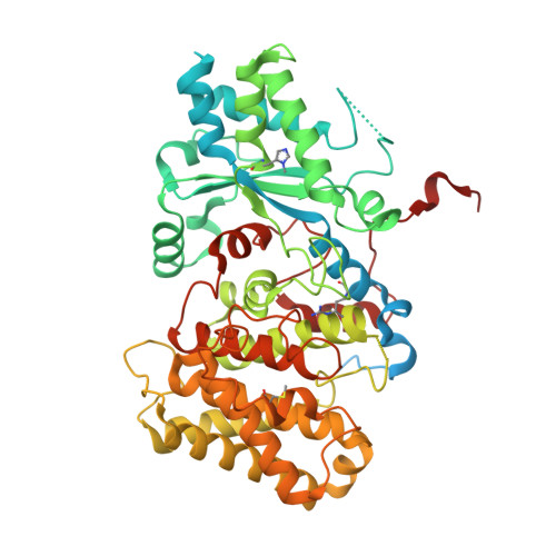
Methyl-coenzyme M reductase subunit alpha
A, B
570Methanosarcina acetivorans C2AMutation(s): 0 
EC: 2.8.4.1
UniProt
Find proteins for Q8THH1 (Methanosarcina acetivorans (strain ATCC 35395 / DSM 2834 / JCM 12185 / C2A))
Explore Q8THH1 
Go to UniProtKB:  Q8THH1
Entity Groups  
Sequence Clusters30% Identity50% Identity70% Identity90% Identity95% Identity100% Identity
UniProt GroupQ8THH1
Sequence Annotations
Expand
  • Reference Sequence
Find similar proteins by:  (by identity cutoff)  |  3D Structure
Entity ID: 2
MoleculeChains Sequence LengthOrganismDetailsImage
Methyl-coenzyme M reductase subunit beta
C, D
434Methanosarcina acetivorans C2AMutation(s): 0 
EC: 2.8.4.1
UniProt
Find proteins for Q8THG7 (Methanosarcina acetivorans (strain ATCC 35395 / DSM 2834 / JCM 12185 / C2A))
Explore Q8THG7 
Go to UniProtKB:  Q8THG7
Entity Groups  
Sequence Clusters30% Identity50% Identity70% Identity90% Identity95% Identity100% Identity
UniProt GroupQ8THG7
Sequence Annotations
Expand
  • Reference Sequence
Find similar proteins by:  (by identity cutoff)  |  3D Structure
Entity ID: 3
MoleculeChains Sequence LengthOrganismDetailsImage
Methyl-coenzyme M reductase subunit gamma
E, F
248Methanosarcina acetivorans C2AMutation(s): 0 
EC: 2.8.4.1
UniProt
Find proteins for Q8THH0 (Methanosarcina acetivorans (strain ATCC 35395 / DSM 2834 / JCM 12185 / C2A))
Explore Q8THH0 
Go to UniProtKB:  Q8THH0
Entity Groups  
Sequence Clusters30% Identity50% Identity70% Identity90% Identity95% Identity100% Identity
UniProt GroupQ8THH0
Sequence Annotations
Expand
  • Reference Sequence
Find similar proteins by:  (by identity cutoff)  |  3D Structure
Entity ID: 4
MoleculeChains Sequence LengthOrganismDetailsImage
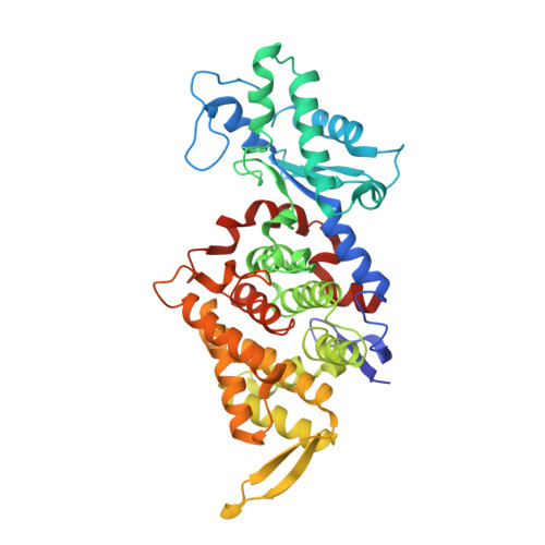
Methyl coenzyme M reductase, subunit DG [auth X]193Methanosarcina acetivorans C2AMutation(s): 0 
Gene Names: mcrD
UniProt
Find proteins for Q8THG8 (Methanosarcina acetivorans (strain ATCC 35395 / DSM 2834 / JCM 12185 / C2A))
Explore Q8THG8 
Go to UniProtKB:  Q8THG8
Entity Groups  
Sequence Clusters30% Identity50% Identity70% Identity90% Identity95% Identity100% Identity
UniProt GroupQ8THG8
Sequence Annotations
Expand
  • Reference Sequence
Small Molecules
Modified Residues  3 Unique
IDChains TypeFormula2D DiagramParent
AGM
Query on AGM
A, B
L-PEPTIDE LINKINGC7 H17 N4 O2ARG
MHS
Query on MHS
A, B
L-PEPTIDE LINKINGC7 H11 N3 O2HIS
SMC
Query on SMC
A, B
L-PEPTIDE LINKINGC4 H9 N O2 SCYS
Experimental Data & Validation

Experimental Data

  • Method: ELECTRON MICROSCOPY
  • Resolution: 3.10 Å
  • Aggregation State: PARTICLE 
  • Reconstruction Method: SINGLE PARTICLE 

Structure Validation

View Full Validation Report


Entry History & Funding Information

Deposition Data


Funding OrganizationLocationGrant Number
Other privateArnold and Mabel Beckman Foundation
David and Lucile Packard FoundationUnited States--
Simons FoundationUnited States822981
Other privateRose Hills Innovator Program
Other privateSearle Scholars Program
Chan Zuckerberg InitiativeUnited States--
Other privateMiller Institute for Basic Research in Science

Revision History  (Full details and data files)

  • Version 1.0: 2023-06-28
    Type: Initial release