8A1M | pdb_00008a1m

Crystal structure of the transpeptidase LdtMt2 from Mycobacterium tuberculosis in complex with maleimide analogue 4


Experimental Data Snapshot

  • Method: X-RAY DIFFRACTION
  • Resolution: 2.30 Å
  • R-Value Free: 
    0.249 (Depositor), 0.250 (DCC) 
  • R-Value Work: 
    0.218 (Depositor), 0.220 (DCC) 
  • R-Value Observed: 
    0.220 (Depositor) 

Starting Model: experimental
View more details

wwPDB Validation   3D Report Full Report


Ligand Structure Quality Assessment 

Created with Raphaël 2.3.0Worse 01 BetterLigand structure goodness of fit to experimental dataBest fitted KULClick on this verticalbar to view details

This is version 1.3 of the entry. See complete history


Literature

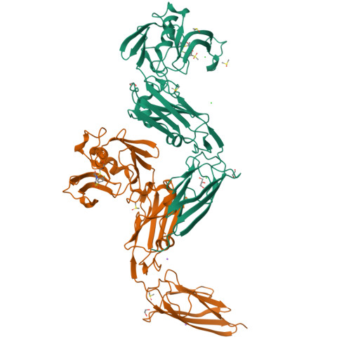
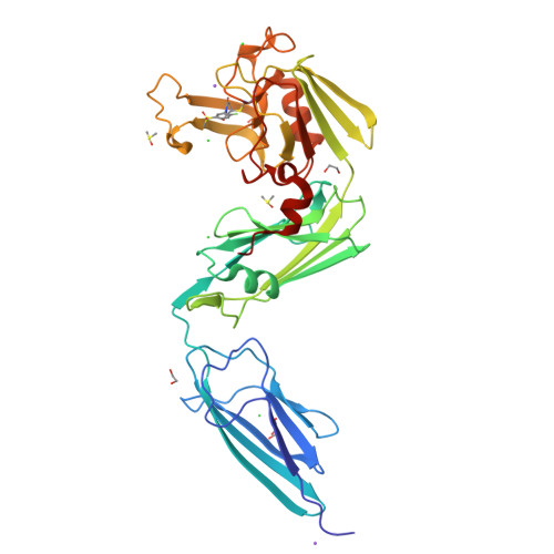
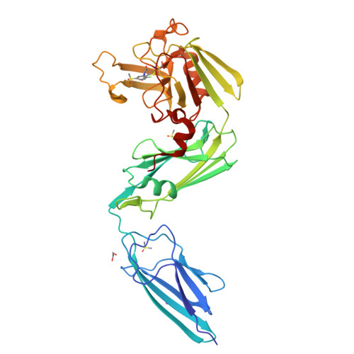
High-throughput screen with the l,d-transpeptidase Ldt Mt2 of Mycobacterium tuberculosis reveals novel classes of covalently reacting inhibitors.

de Munnik, M.Lang, P.A.De Dios Anton, F.Cacho, M.Bates, R.H.Brem, J.Rodriguez Miquel, B.Schofield, C.J.

(2023) Chem Sci 14: 7262-7278

  • DOI: https://doi.org/10.1039/d2sc06858c
  • Primary Citation of Related Structures:  
    8A1J, 8A1K, 8A1L, 8A1M, 8A1N, 8A1O, 8AHO

  • PubMed Abstract: 

    Disruption of bacterial cell wall biosynthesis in Mycobacterium tuberculosis is a promising target for treating tuberculosis. The l,d-transpeptidase Ldt Mt2 , which is responsible for the formation of 3 → 3 cross-links in the cell wall peptidoglycan, has been identified as essential for M. tuberculosis virulence. We optimised a high-throughput assay for Ldt Mt2 , and screened a targeted library of ∼10 000 electrophilic compounds. Potent inhibitor classes were identified, including established ( e.g. , β-lactams) and unexplored covalently reacting electrophilic groups ( e.g. , cyanamides). Protein-observed mass spectrometric studies reveal most classes to react covalently and irreversibly with the Ldt Mt2 catalytic cysteine (Cys354). Crystallographic analyses of seven representative inhibitors reveal induced fit involving a loop enclosing the Ldt Mt2 active site. Several of the identified compounds have a bactericidal effect on M. tuberculosis within macrophages, one with an MIC 50 value of ∼1 μM. The results provide leads for the development of new covalently reaction inhibitors of Ldt Mt2 and other nucleophilic cysteine enzymes.


  • Organizational Affiliation

    Chemistry Research Laboratory, Department of Chemistry, the Ineos Oxford Institute of Antimicrobial Research, University of Oxford 12 Mansfield Road Oxford OX1 3TA UK christopher.schofield@chem.ox.ac.uk.


Macromolecules
Find similar proteins by:  (by identity cutoff)  |  3D Structure
Entity ID: 1
MoleculeChains Sequence LengthOrganismDetailsImage
L,D-transpeptidase 2
A, B
355Mycobacterium tuberculosisMutation(s): 0 
Gene Names: ldtBMT2594V735_02606
EC: 2.3.2
UniProt
Find proteins for I6Y9J2 (Mycobacterium tuberculosis (strain ATCC 25618 / H37Rv))
Explore I6Y9J2 
Go to UniProtKB:  I6Y9J2
Entity Groups  
Sequence Clusters30% Identity50% Identity70% Identity90% Identity95% Identity100% Identity
UniProt GroupI6Y9J2
Sequence Annotations
Expand
  • Reference Sequence
Small Molecules
Ligands 6 Unique
IDChains Name / Formula / InChI Key2D Diagram3D Interactions
KUL (Subject of Investigation/LOI)
Query on KUL

Download Ideal Coordinates CCD File 
C [auth A],
P [auth B]
1-(2-fluoranyl-5-methylsulfonyl-phenyl)pyrrolidine-2,5-dione
C11 H10 F N O4 S
RISNMQBAJMCDFN-UHFFFAOYSA-N
GOL
Query on GOL

Download Ideal Coordinates CCD File 
G [auth A]GLYCEROL
C3 H8 O3
PEDCQBHIVMGVHV-UHFFFAOYSA-N
DMS
Query on DMS

Download Ideal Coordinates CCD File 
E [auth A],
F [auth A],
I [auth A],
Q [auth B],
R [auth B]
DIMETHYL SULFOXIDE
C2 H6 O S
IAZDPXIOMUYVGZ-UHFFFAOYSA-N
EDO
Query on EDO

Download Ideal Coordinates CCD File 
D [auth A],
H [auth A],
S [auth B]
1,2-ETHANEDIOL
C2 H6 O2
LYCAIKOWRPUZTN-UHFFFAOYSA-N
CL
Query on CL

Download Ideal Coordinates CCD File 
L [auth A],
M [auth A],
N [auth A],
O [auth A]
CHLORIDE ION
Cl
VEXZGXHMUGYJMC-UHFFFAOYSA-M
NA
Query on NA

Download Ideal Coordinates CCD File 
J [auth A],
K [auth A],
T [auth B]
SODIUM ION
Na
FKNQFGJONOIPTF-UHFFFAOYSA-N
Experimental Data & Validation

Experimental Data

  • Method: X-RAY DIFFRACTION
  • Resolution: 2.30 Å
  • R-Value Free:  0.249 (Depositor), 0.250 (DCC) 
  • R-Value Work:  0.218 (Depositor), 0.220 (DCC) 
  • R-Value Observed: 0.220 (Depositor) 
Space Group: P 1 21 1
Unit Cell:
Length ( Å )Angle ( ˚ )
a = 61.7α = 90
b = 95.07β = 92.22
c = 75.45γ = 90
Software Package:
Software NamePurpose
PHENIXrefinement
XDSdata reduction
xia2data scaling
PHASERphasing
PDB_EXTRACTdata extraction

Structure Validation

View Full Validation Report


Ligand Structure Quality Assessment 

Created with Raphaël 2.3.0Worse 01 BetterLigand structure goodness of fit to experimental dataBest fitted KULClick on this verticalbar to view details

Entry History & Funding Information

Deposition Data


Funding OrganizationLocationGrant Number
Biotechnology and Biological Sciences Research Council (BBSRC)United Kingdom--

Revision History  (Full details and data files)

  • Version 1.0: 2023-06-14
    Type: Initial release
  • Version 1.1: 2023-08-02
    Changes: Database references
  • Version 1.2: 2024-02-07
    Changes: Data collection, Refinement description
  • Version 1.3: 2024-10-16
    Changes: Structure summary