7R4P

Structure of human hydroxyacid oxidase 1 bound with 6-amino-1-benzyl-5-(methylamino)pyrimidine-2,4(1H,3H)-dione


Experimental Data Snapshot

  • Method: X-RAY DIFFRACTION
  • Resolution: 1.37 Å
  • R-Value Free: 
    0.196 (Depositor), 0.200 (DCC) 
  • R-Value Work: 
    0.180 (Depositor), 0.180 (DCC) 
  • R-Value Observed: 
    0.181 (Depositor) 

Starting Model: experimental
View more details

wwPDB Validation   3D Report Full Report


Ligand Structure Quality Assessment 

Created with Raphaël 2.3.0Worse 01 BetterLigand structure goodness of fit to experimental dataBest fitted I2ZClick on this verticalbar to view details

This is version 1.1 of the entry. See complete history


Literature

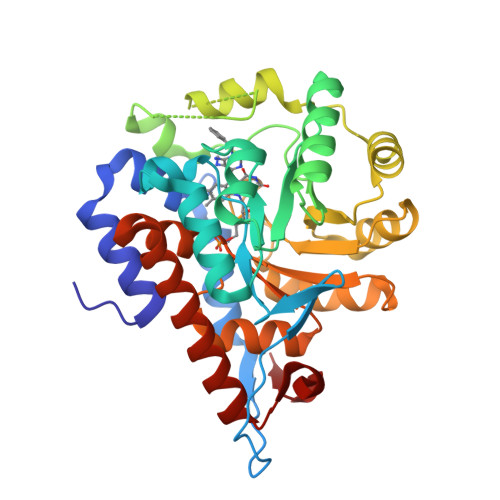
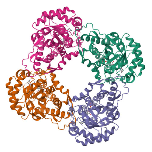
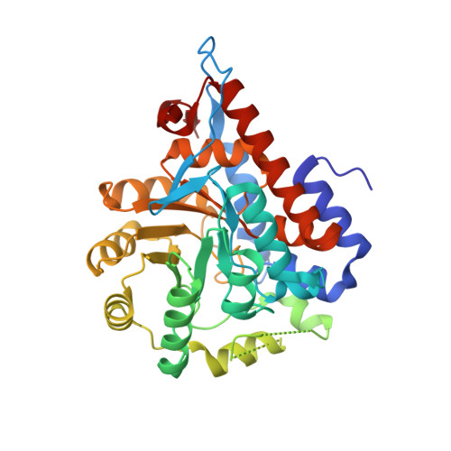
Structure of human hydroxyacid oxidase 1 bound with 5-bromo-N-methyl-1H-indazole-3-carboxamide

Mackinnon, S.Bezerra, G.A.Krojer, T.Bradley, A.R.Talon, R.Brandeo-Neto, J.Douangamath, A.von Delft, F.Arrowsmith, C.H.Edwards, A.Bountra, C.Oppermann, U.Brennan, P.E.Yue, W.W.

To be published.

Macromolecules
Find similar proteins by:  (by identity cutoff)  |  3D Structure
Entity ID: 1
MoleculeChains Sequence LengthOrganismDetailsImage
Hydroxyacid oxidase 1362Homo sapiensMutation(s): 0 
Gene Names: HAO1GOX1HAOX1
EC: 1.1.3.15 (PDB Primary Data), 1.2.3.5 (UniProt)
UniProt & NIH Common Fund Data Resources
Find proteins for Q9UJM8 (Homo sapiens)
Explore Q9UJM8 
Go to UniProtKB:  Q9UJM8
PHAROS:  Q9UJM8
GTEx:  ENSG00000101323 
Entity Groups  
Sequence Clusters30% Identity50% Identity70% Identity90% Identity95% Identity100% Identity
UniProt GroupQ9UJM8
Sequence Annotations
Expand
  • Reference Sequence
Small Molecules
Ligands 2 Unique
IDChains Name / Formula / InChI Key2D Diagram3D Interactions
FMN
Query on FMN

Download Ideal Coordinates CCD File 
B [auth A]FLAVIN MONONUCLEOTIDE
C17 H21 N4 O9 P
FVTCRASFADXXNN-SCRDCRAPSA-N
I2Z (Subject of Investigation/LOI)
Query on I2Z

Download Ideal Coordinates CCD File 
C [auth A]6-amino-1-benzyl-5-(methylamino)pyrimidine-2,4(1H,3H)-dione
C12 H16 N4 O2
ROGMXQYZAWKCLY-ZJUUUORDSA-N
Experimental Data & Validation

Experimental Data

  • Method: X-RAY DIFFRACTION
  • Resolution: 1.37 Å
  • R-Value Free:  0.196 (Depositor), 0.200 (DCC) 
  • R-Value Work:  0.180 (Depositor), 0.180 (DCC) 
  • R-Value Observed: 0.181 (Depositor) 
Space Group: I 4
Unit Cell:
Length ( Å )Angle ( ˚ )
a = 97.13α = 90
b = 97.13β = 90
c = 80.84γ = 90
Software Package:
Software NamePurpose
REFMACrefinement
Aimlessdata scaling
PDB_EXTRACTdata extraction
XDSdata reduction
PHASERphasing

Structure Validation

View Full Validation Report


Ligand Structure Quality Assessment 

Created with Raphaël 2.3.0Worse 01 BetterLigand structure goodness of fit to experimental dataBest fitted I2ZClick on this verticalbar to view details

Entry History & Funding Information

Deposition Data


Funding OrganizationLocationGrant Number
Wellcome TrustUnited Kingdom092809/Z/10/Z

Revision History  (Full details and data files)

  • Version 1.0: 2022-05-04
    Type: Initial release
  • Version 1.1: 2024-01-31
    Changes: Data collection, Refinement description