7LUN | pdb_00007lun

Human PARP14 (ARTD8), catalytic fragment in complex with RBN011980


Experimental Data Snapshot

  • Method: X-RAY DIFFRACTION
  • Resolution: 2.57 Å
  • R-Value Free: 
    0.278 (Depositor), 0.280 (DCC) 
  • R-Value Work: 
    0.232 (Depositor), 0.240 (DCC) 
  • R-Value Observed: 
    0.235 (Depositor) 

Starting Model: experimental
View more details

wwPDB Validation   3D Report Full Report


Ligand Structure Quality Assessment 

Created with Raphaël 2.3.0Worse 01 BetterLigand structure goodness of fit to experimental dataBest fitted YFGClick on this verticalbar to view details

This is version 1.3 of the entry. See complete history


Literature

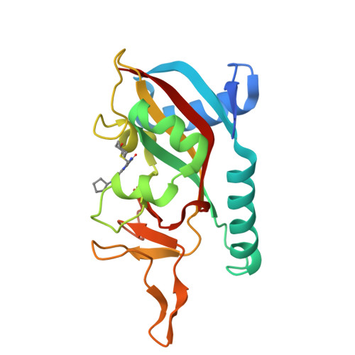
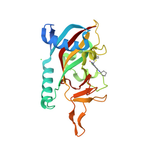
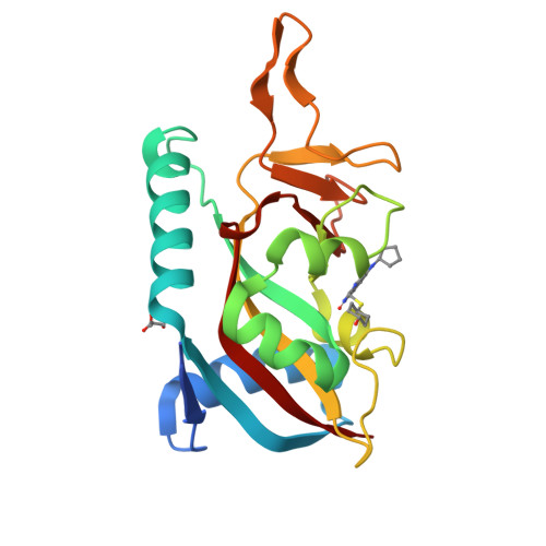
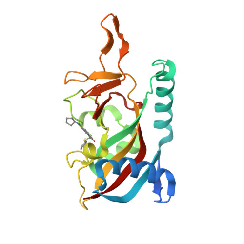
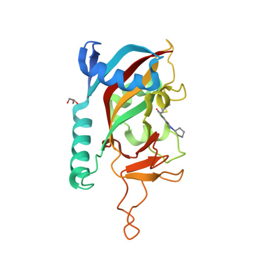
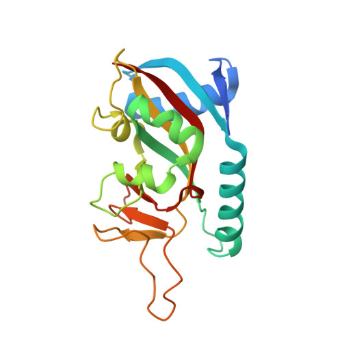
Targeted Degradation of PARP14 Using a Heterobifunctional Small Molecule.

Wigle, T.J.Ren, Y.Molina, J.R.Blackwell, D.J.Schenkel, L.B.Swinger, K.K.Kuplast-Barr, K.Majer, C.R.Church, W.D.Lu, A.Z.Mo, J.Abo, R.Cheung, A.Dorsey, B.W.Niepel, M.Perl, N.R.Vasbinder, M.M.Keilhack, H.Kuntz, K.W.

(2021) Chembiochem 22: 2107-2110

  • DOI: https://doi.org/10.1002/cbic.202100047
  • Primary Citation of Related Structures:  
    7L9Y, 7LUN

  • PubMed Abstract: 

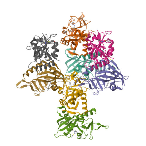
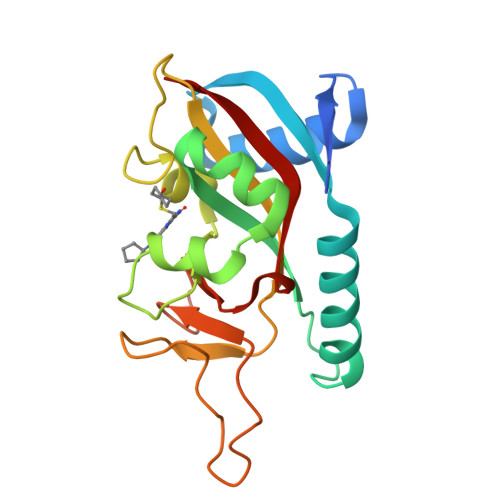
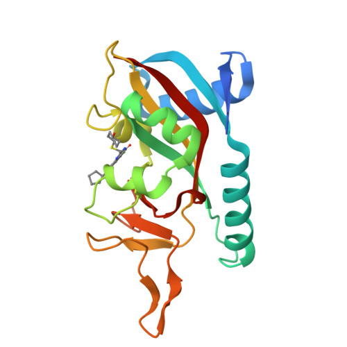
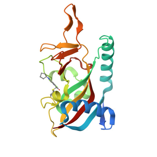
    PARP14 is an interferon-stimulated gene that is overexpressed in multiple tumor types, influencing pro-tumor macrophage polarization as well as suppressing the antitumor inflammation response by modulating IFN-γ and IL-4 signaling. PARP14 is a 203 kDa protein that possesses a catalytic domain responsible for the transfer of mono-ADP-ribose to its substrates. PARP14 also contains three macrodomains and a WWE domain which are binding modules for mono-ADP-ribose and poly-ADP-ribose, respectively, in addition to two RNA recognition motifs. Catalytic inhibitors of PARP14 have been shown to reverse IL-4 driven pro-tumor gene expression in macrophages, however it is not clear what roles the non-enzymatic biomolecular recognition motifs play in PARP14-driven immunology and inflammation. To further understand this, we have discovered a heterobifunctional small molecule designed based on a catalytic inhibitor of PARP14 that binds in the enzyme's NAD + -binding site and recruits cereblon to ubiquitinate it and selectively target it for degradation.


  • Organizational Affiliation

    Ribon Therapeutics, 35 Cambridgepark Dr., Suite 300, Cambridge, MA 02140, USA.


Macromolecules
Find similar proteins by:  (by identity cutoff)  |  3D Structure
Entity ID: 1
MoleculeChains Sequence LengthOrganismDetailsImage
Protein mono-ADP-ribosyltransferase PARP14
A, B, C, D, E
194Homo sapiensMutation(s): 0 
Gene Names: PARP14BAL2KIAA1268
EC: 2.4.2
UniProt & NIH Common Fund Data Resources
Find proteins for Q460N5 (Homo sapiens)
Explore Q460N5 
Go to UniProtKB:  Q460N5
PHAROS:  Q460N5
GTEx:  ENSG00000173193 
Entity Groups  
Sequence Clusters30% Identity50% Identity70% Identity90% Identity95% Identity100% Identity
UniProt GroupQ460N5
Sequence Annotations
Expand
  • Reference Sequence
Small Molecules
Ligands 3 Unique
IDChains Name / Formula / InChI Key2D Diagram3D Interactions
YFG (Subject of Investigation/LOI)
Query on YFG

Download Ideal Coordinates CCD File 
I [auth A]
J [auth B]
K [auth C]
L [auth D]
M [auth E]
7-(cyclopentylamino)-5-fluoro-2-{[(trans-4-hydroxycyclohexyl)sulfanyl]methyl}quinazolin-4(3H)-one
C20 H26 F N3 O2 S
FUHIIFURQIAPDZ-SHTZXODSSA-N
GOL
Query on GOL

Download Ideal Coordinates CCD File 
P [auth F],
S [auth H]
GLYCEROL
C3 H8 O3
PEDCQBHIVMGVHV-UHFFFAOYSA-N
CL
Query on CL

Download Ideal Coordinates CCD File 
N [auth E]CHLORIDE ION
Cl
VEXZGXHMUGYJMC-UHFFFAOYSA-M
Binding Affinity Annotations 
IDSourceBinding Affinity
YFG BindingDB:  7LUN IC50: min: 1000, max: 1000 (nM) from 2 assay(s)
Experimental Data & Validation

Experimental Data

  • Method: X-RAY DIFFRACTION
  • Resolution: 2.57 Å
  • R-Value Free:  0.278 (Depositor), 0.280 (DCC) 
  • R-Value Work:  0.232 (Depositor), 0.240 (DCC) 
  • R-Value Observed: 0.235 (Depositor) 
Space Group: P 1 21 1
Unit Cell:
Length ( Å )Angle ( ˚ )
a = 65.99α = 90
b = 153.66β = 108.7
c = 87.46γ = 90
Software Package:
Software NamePurpose
REFMACrefinement
XDSdata reduction
Aimlessdata scaling
PHASERphasing
PDB_EXTRACTdata extraction

Structure Validation

View Full Validation Report


Ligand Structure Quality Assessment 

Created with Raphaël 2.3.0Worse 01 BetterLigand structure goodness of fit to experimental dataBest fitted YFGClick on this verticalbar to view details

Entry History 

Deposition Data

Revision History  (Full details and data files)

  • Version 1.0: 2021-04-21
    Type: Initial release
  • Version 1.1: 2021-06-23
    Changes: Database references
  • Version 1.2: 2023-10-18
    Changes: Data collection, Database references, Refinement description
  • Version 1.3: 2024-10-23
    Changes: Structure summary