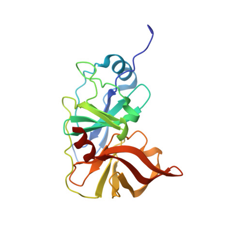
7L7P

Crystal structure of HCV NS3/4A D168A protease in complex with CH-24

  • Classification: HYDROLASE/INHIBITOR
  • Organism(s): Hepacivirus hominis
  • Expression System: Escherichia coli BL21(DE3)
  • Mutation(s): Yes 

  • Deposited: 2020-12-29 Released: 2021-09-01 
  • Deposition Author(s): Zephyr, J., Schiffer, C.A.
  • Funding Organization(s): National Institutes of Health/National Institute Of Allergy and Infectious Diseases (NIH/NIAID), National Institutes of Health/National Institute of General Medical Sciences (NIH/NIGMS)

Experimental Data Snapshot

  • Method: X-RAY DIFFRACTION
  • Resolution: 1.58 Å
  • R-Value Free: 0.184 
  • R-Value Work: 0.163 
  • R-Value Observed: 0.164 

Starting Model: experimental
View more details

wwPDB Validation   3D Report Full Report


Ligand Structure Quality Assessment 


This is version 1.2 of the entry. See complete history


Literature

Discovery of Quinoxaline-Based P1-P3 Macrocyclic NS3/4A Protease Inhibitors with Potent Activity against Drug-Resistant Hepatitis C Virus Variants.

Nageswara Rao, D.Zephyr, J.Henes, M.Chan, E.T.Matthew, A.N.Hedger, A.K.Conway, H.L.Saeed, M.Newton, A.Petropoulos, C.J.Huang, W.Kurt Yilmaz, N.Schiffer, C.A.Ali, A.

(2021) J Med Chem 64: 11972-11989

  • DOI: https://doi.org/10.1021/acs.jmedchem.1c00554
  • Primary Citation of Related Structures:  
    7L7L, 7L7N, 7L7O, 7L7P

  • PubMed Abstract: 

    The three pan-genotypic HCV NS3/4A protease inhibitors (PIs) currently in clinical use-grazoprevir, glecaprevir, and voxilaprevir-are quinoxaline-based P2-P4 macrocycles and thus exhibit similar resistance profiles. Using our quinoxaline-based P1-P3 macrocyclic lead compounds as an alternative chemical scaffold, we explored structure-activity relationships (SARs) at the P2 and P4 positions to develop pan-genotypic PIs that avoid drug resistance. A structure-guided strategy was used to design and synthesize two series of compounds with different P2 quinoxalines in combination with diverse P4 groups of varying sizes and shapes, with and without fluorine substitutions. Our SAR data and cocrystal structures revealed the interplay between the P2 and P4 groups, which influenced inhibitor binding and the overall resistance profile. Optimizing inhibitor interactions in the S4 pocket led to PIs with excellent antiviral activity against clinically relevant PI-resistant HCV variants and genotype 3, providing potential pan-genotypic inhibitors with improved resistance profiles.


  • Organizational Affiliation

    Department of Biochemistry and Molecular Pharmacology, University of Massachusetts Medical School, Worcester, Massachusetts 01605, United States.


Macromolecules
Find similar proteins by:  (by identity cutoff)  |  3D Structure
Entity ID: 1
MoleculeChains Sequence LengthOrganismDetailsImage
NS3/4A protease200Hepacivirus hominisMutation(s): 1 
UniProt
Find proteins for A0A0B4WYC6 (Hepacivirus hominis)
Explore A0A0B4WYC6 
Go to UniProtKB:  A0A0B4WYC6
Entity Groups  
Sequence Clusters30% Identity50% Identity70% Identity90% Identity95% Identity100% Identity
UniProt GroupA0A0B4WYC6
Sequence Annotations
Expand
  • Reference Sequence
Small Molecules
Ligands 4 Unique
IDChains Name / Formula / InChI Key2D Diagram3D Interactions
XT4 (Subject of Investigation/LOI)
Query on XT4

Download Ideal Coordinates CCD File 
C [auth A]tert-butyl [(2R,6S,12Z,13aS,14aR,16aS)-2-{[6-methoxy-3-(trifluoromethyl)quinoxalin-2-yl]oxy}-14a-{[(1-methylcyclopropyl)sulfonyl]carbamoyl}-5,16-dioxo-1,2,3,5,6,7,8,9,10,11,13a,14,14a,15,16,16a-hexadecahydrocyclopropa[e]pyrrolo[1,2-a][1,4]diazacyclopentadecin-6-yl]carbamate
C37 H47 F3 N6 O9 S
IYYDCFFQTJJOBE-NTPKMKHWSA-N
SO4
Query on SO4

Download Ideal Coordinates CCD File 
H [auth A]SULFATE ION
O4 S
QAOWNCQODCNURD-UHFFFAOYSA-L
ZN
Query on ZN

Download Ideal Coordinates CCD File 
B [auth A]ZINC ION
Zn
PTFCDOFLOPIGGS-UHFFFAOYSA-N
EDO
Query on EDO

Download Ideal Coordinates CCD File 
D [auth A],
E [auth A],
F [auth A],
G [auth A]
1,2-ETHANEDIOL
C2 H6 O2
LYCAIKOWRPUZTN-UHFFFAOYSA-N
Experimental Data & Validation

Experimental Data

  • Method: X-RAY DIFFRACTION
  • Resolution: 1.58 Å
  • R-Value Free: 0.184 
  • R-Value Work: 0.163 
  • R-Value Observed: 0.164 
  • Space Group: P 21 21 21
Unit Cell:
Length ( Å )Angle ( ˚ )
a = 55.062α = 90
b = 60.042β = 90
c = 58.808γ = 90
Software Package:
Software NamePurpose
PHENIXrefinement
PHASERphasing
HKL-3000data scaling
Cootmodel building
HKL-3000data reduction

Structure Validation

View Full Validation Report


Ligand Structure Quality Assessment 


Entry History & Funding Information

Deposition Data


Funding OrganizationLocationGrant Number
National Institutes of Health/National Institute Of Allergy and Infectious Diseases (NIH/NIAID)United StatesR01 AI085051
National Institutes of Health/National Institute of General Medical Sciences (NIH/NIGMS)United StatesF31 GM131635

Revision History  (Full details and data files)

  • Version 1.0: 2021-09-01
    Type: Initial release
  • Version 1.1: 2021-09-08
    Changes: Database references
  • Version 1.2: 2023-10-18
    Changes: Data collection, Refinement description