7BD0

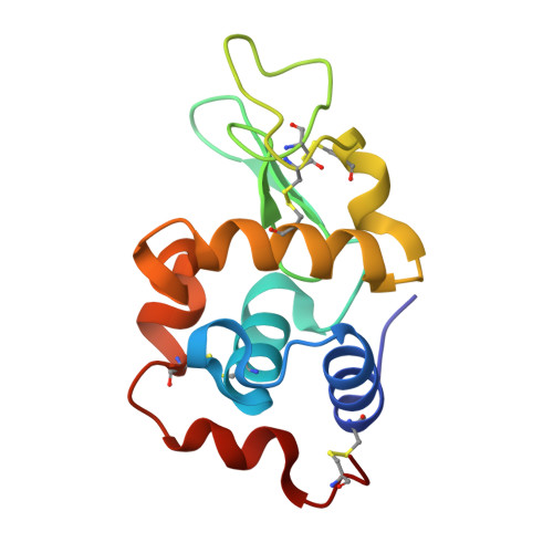
The adduct of NAMI-A with Hen Egg White Lysozyme at 26 hours.


Experimental Data Snapshot

  • Method: X-RAY DIFFRACTION
  • Resolution: 1.06 Å
  • R-Value Free: 0.185 
  • R-Value Work: 0.164 
  • R-Value Observed: 0.165 

Starting Model: experimental
View more details

wwPDB Validation   3D Report Full Report


This is version 1.2 of the entry. See complete history


Literature

Insights into the Protein Ruthenation Mechanism by Antimetastatic Metallodrugs: High-Resolution X-ray Structures of the Adduct Formed between Hen Egg-White Lysozyme and NAMI-A at Various Time Points.

Chiniadis, L.Giastas, P.Bratsos, I.Papakyriakou, A.

(2021) Inorg Chem 60: 10729-10737

  • DOI: https://doi.org/10.1021/acs.inorgchem.1c01441
  • Primary Citation of Related Structures:  
    7BCU, 7BCX, 7BD0, 7BDM

  • PubMed Abstract: 

    The pharmacological profile of medicinally relevant Ru(III) coordination compounds has been ascribed to their interactions with proteins, as several studies have provided evidence that DNA is not the primary target. In this regard, numerous spectroscopic and crystallographic studies have indicated that the Ru(III) ligands play an important role in determining the metal binding site, acting as the recognition element in the early stages of the protein-complex formation. Herein, we present a series of near-atomic-resolution X-ray crystal structures of the adducts formed between the antimetastatic metallodrug imidazolium trans -[tetrachlorido( S -dimethyl sufoxide)(1 H -imidazole)ruthenate(III)] ( NAMI-A ) and hen egg-white lysozyme (HEWL). These structures elucidate a series of binding events starting from the noncovalent interaction of intact NAMI-A ions with HEWL (1.5 h), followed by the stepwise exchange of all Ru ligands except for 1 H -imidazole (26 h) to the final "ruthenated" protein comprising one aquated Ru ion coordinated to histidine-15 of HEWL (98 h). Our structural data clearly support a two-step mechanism of protein ruthenation, illustrating the ligand-mediated recognition step of the process.


  • Organizational Affiliation

    Department of Neurobiology, Hellenic Pasteur Institute, 11521 Athens, Greece.


Macromolecules
Find similar proteins by:  (by identity cutoff)  |  3D Structure
Entity ID: 1
MoleculeChains Sequence LengthOrganismDetailsImage
Lysozyme129Gallus gallusMutation(s): 0 
EC: 3.2.1.17
UniProt
Find proteins for P00698 (Gallus gallus)
Explore P00698 
Go to UniProtKB:  P00698
Entity Groups  
Sequence Clusters30% Identity50% Identity70% Identity90% Identity95% Identity100% Identity
UniProt GroupP00698
Sequence Annotations
Expand
  • Reference Sequence
Small Molecules
Ligands 5 Unique
IDChains Name / Formula / InChI Key2D Diagram3D Interactions
RU (Subject of Investigation/LOI)
Query on RU

Download Ideal Coordinates CCD File 
H [auth A],
K [auth A],
N [auth A]
RUTHENIUM ION
Ru
BPEVHDGLPIIAGH-UHFFFAOYSA-N
IMD
Query on IMD

Download Ideal Coordinates CCD File 
G [auth A],
J [auth A],
M [auth A]
IMIDAZOLE
C3 H5 N2
RAXXELZNTBOGNW-UHFFFAOYSA-O
EDO
Query on EDO

Download Ideal Coordinates CCD File 
B [auth A],
C [auth A]
1,2-ETHANEDIOL
C2 H6 O2
LYCAIKOWRPUZTN-UHFFFAOYSA-N
CL
Query on CL

Download Ideal Coordinates CCD File 
I [auth A],
L [auth A]
CHLORIDE ION
Cl
VEXZGXHMUGYJMC-UHFFFAOYSA-M
NA
Query on NA

Download Ideal Coordinates CCD File 
D [auth A],
E [auth A],
F [auth A]
SODIUM ION
Na
FKNQFGJONOIPTF-UHFFFAOYSA-N
Experimental Data & Validation

Experimental Data

  • Method: X-RAY DIFFRACTION
  • Resolution: 1.06 Å
  • R-Value Free: 0.185 
  • R-Value Work: 0.164 
  • R-Value Observed: 0.165 
  • Space Group: P 43 21 2
Unit Cell:
Length ( Å )Angle ( ˚ )
a = 78.298α = 90
b = 78.298β = 90
c = 37.571γ = 90
Software Package:
Software NamePurpose
PHENIXrefinement
XDSdata reduction
XSCALEdata scaling
PHASERphasing

Structure Validation

View Full Validation Report


Entry History 

Deposition Data

Revision History  (Full details and data files)

  • Version 1.0: 2021-07-28
    Type: Initial release
  • Version 1.1: 2024-01-31
    Changes: Data collection, Database references, Refinement description
  • Version 1.2: 2024-11-20
    Changes: Structure summary