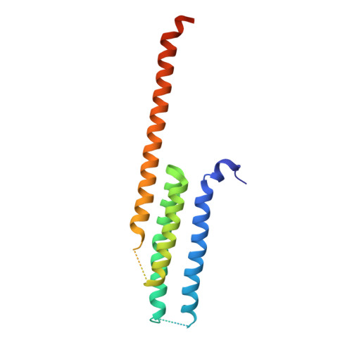
6VGI

Crystal Structures of FLAP bound to MK-866


Experimental Data Snapshot

  • Method: X-RAY DIFFRACTION
  • Resolution: 2.61 Å
  • R-Value Free: 0.276 
  • R-Value Work: 0.259 
  • R-Value Observed: 0.259 

Starting Model: experimental
View more details

wwPDB Validation   3D Report Full Report

Currently 6VGI does not have a validation slider image.


This is version 1.3 of the entry. See complete history


Literature

Structure-based, multi-targeted drug discovery approach to eicosanoid inhibition: Dual inhibitors of mPGES-1 and 5-lipoxygenase activating protein (FLAP).

Ho, J.D.Lee, M.R.Rauch, C.T.Aznavour, K.Park, J.S.Luz, J.G.Antonysamy, S.Condon, B.Maletic, M.Zhang, A.Hickey, M.J.Hughes, N.E.Chandrasekhar, S.Sloan, A.V.Gooding, K.Harvey, A.Yu, X.P.Kahl, S.D.Norman, B.H.

(2020) Biochim Biophys Acta Gen Subj 1865: 129800-129800

  • DOI: https://doi.org/10.1016/j.bbagen.2020.129800
  • Primary Citation of Related Structures:  
    6VGC, 6VGI, 6VL4

  • PubMed Abstract: 

    Due to the importance of both prostaglandins (PGs) and leukotrienes (LTs) as pro-inflammatory mediators, and the potential for eicosanoid shunting in the presence of pathway target inhibitors, we have investigated an approach to inhibiting the formation of both PGs and LTs as part of a multi-targeted drug discovery effort. We generated ligand-protein X-ray crystal structures of known inhibitors of microsomal prostaglandin E2 synthase-1 (mPGES-1) and the 5-Lipoxygenase Activating Protein (FLAP), with their respective proteins, to understand the overlapping pharmacophores. We subsequently used molecular modeling and structure-based drug design (SBDD) to identify hybrid structures intended to inhibit both targets. This work enabled the preparation of compounds 4 and 5, which showed potent in vitro inhibition of both targets. Our findings enhance the structural understanding of mPGES-1 and FLAP's unique ligand binding pockets and should accelerate the discovery of additional dual inhibitors for these two important integral membrane protein drug targets.


  • Organizational Affiliation

    Lilly Biotechnology Center, San Diego, CA 92121, USA. Electronic address: [email protected].


Macromolecules
Find similar proteins by:  (by identity cutoff)  |  3D Structure
Entity ID: 1
MoleculeChains Sequence LengthOrganismDetailsImage
5-lipoxygenase-activating protein
A, B, C, D, E
A, B, C, D, E, F
171Homo sapiensMutation(s): 0 
Gene Names: ALOX5APFLAP
Membrane Entity: Yes 
UniProt & NIH Common Fund Data Resources
Find proteins for P20292 (Homo sapiens)
Explore P20292 
Go to UniProtKB:  P20292
PHAROS:  P20292
GTEx:  ENSG00000132965 
Entity Groups  
Sequence Clusters30% Identity50% Identity70% Identity90% Identity95% Identity100% Identity
UniProt GroupP20292
Sequence Annotations
Expand
  • Reference Sequence
Small Molecules
Ligands 2 Unique
IDChains Name / Formula / InChI Key2D Diagram3D Interactions
QY7 (Subject of Investigation/LOI)
Query on QY7

Download Ideal Coordinates CCD File 
G [auth A]
I [auth B]
J [auth B]
L [auth C]
N [auth E]
G [auth A],
I [auth B],
J [auth B],
L [auth C],
N [auth E],
P [auth F]
3-[3-(tert-butylsulfanyl)-1-[(4-chlorophenyl)methyl]-5-(propan-2-yl)-1H-indol-2-yl]-2,2-dimethylpropanoic acid
C27 H34 Cl N O2 S
QAOAOVKBIIKRNL-UHFFFAOYSA-N
SO4
Query on SO4

Download Ideal Coordinates CCD File 
H [auth A],
K [auth B],
M [auth C],
O [auth E],
Q [auth F]
SULFATE ION
O4 S
QAOWNCQODCNURD-UHFFFAOYSA-L
Binding Affinity Annotations 
IDSourceBinding Affinity
QY7 BindingDB:  6VGI IC50: min: 2.2, max: 1400 (nM) from 13 assay(s)
Experimental Data & Validation

Experimental Data

  • Method: X-RAY DIFFRACTION
  • Resolution: 2.61 Å
  • R-Value Free: 0.276 
  • R-Value Work: 0.259 
  • R-Value Observed: 0.259 
  • Space Group: C 1 2 1
Unit Cell:
Length ( Å )Angle ( ˚ )
a = 214.418α = 90
b = 66.297β = 101.53
c = 109.43γ = 90
Software Package:
Software NamePurpose
REFMACrefinement
PDB_EXTRACTdata extraction
MOSFLMdata reduction
SCALAdata scaling
PHASERphasing

Structure Validation

View Full Validation Report

Currently 6VGI does not have a validation slider image.


Entry History 

Revision History  (Full details and data files)

  • Version 1.0: 2020-12-02
    Type: Initial release
  • Version 1.1: 2020-12-09
    Changes: Database references
  • Version 1.2: 2020-12-16
    Changes: Database references
  • Version 1.3: 2023-10-11
    Changes: Data collection, Database references, Refinement description