6U6V

Crystal structure of human PD-1H / VISTA


Experimental Data Snapshot

  • Method: X-RAY DIFFRACTION
  • Resolution: 1.90 Å
  • R-Value Free: 
    0.250 (Depositor), 0.255 (DCC) 
  • R-Value Work: 
    0.207 (Depositor), 0.214 (DCC) 
  • R-Value Observed: 
    0.210 (Depositor) 

wwPDB Validation   3D Report Full Report


Ligand Structure Quality Assessment 

Created with Raphaël 2.3.0Worse 01 BetterLigand structure goodness of fit to experimental dataBest fitted NAGClick on this verticalbar to view details

This is version 1.4 of the entry. See complete history


Literature

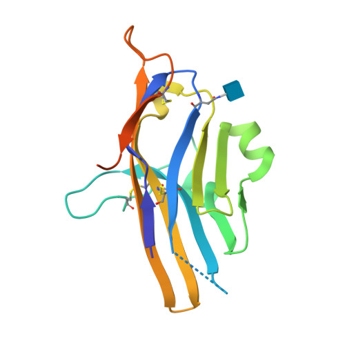
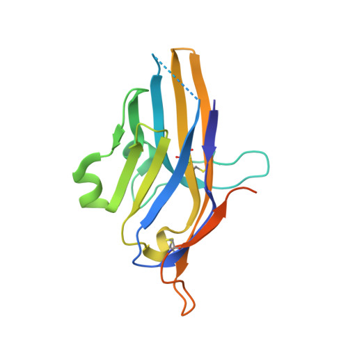
Structural insight into T cell coinhibition by PD-1H (VISTA).

Slater, B.T.Han, X.Chen, L.Xiong, Y.

(2020) Proc Natl Acad Sci U S A 117: 1648-1657

  • DOI: https://doi.org/10.1073/pnas.1908711117
  • Primary Citation of Related Structures:  
    6U6V

  • PubMed Abstract: 

    Programmed death-1 homolog (PD-1H), a CD28/B7 family molecule, coinhibits T cell activation and is an attractive immunotherapeutic target for cancer and inflammatory diseases. The molecular basis of its function, however, is unknown. Bioinformatic analyses indicated that PD-1H has a very long Ig variable region (IgV)-like domain and extraordinarily high histidine content, suggesting that unique structural features may contribute to coinhibitory mechanisms. Here we present the 1.9-Å crystal structure of the human PD-1H extracellular domain. It reveals an elongated CC' loop and a striking concentration of histidine residues, located in the complementarity-determining region-like proximal half of the molecule. We show that surface-exposed histidine clusters are essential for robust inhibition of T cell activation. PD-1H exhibits a noncanonical IgV-like topology including an extra "H" β-strand and "clamping" disulfide, absent in known IgV-like structures, that likely restricts its orientation on the cell surface differently from other IgV-like domains. These results provide important insight into a molecular basis of T cell coinhibition by PD-1H.


  • Organizational Affiliation

    Department of Immunobiology, Yale University, New Haven, CT 06511.


Macromolecules
Find similar proteins by:  (by identity cutoff)  |  3D Structure
Entity ID: 1
MoleculeChains Sequence LengthOrganismDetailsImage
V-type immunoglobulin domain-containing suppressor of T-cell activation174Homo sapiensMutation(s): 0 
Gene Names: VSIRC10orf54SISP1VISTAPP2135UNQ730/PRO1412
UniProt & NIH Common Fund Data Resources
Find proteins for Q9H7M9 (Homo sapiens)
Explore Q9H7M9 
Go to UniProtKB:  Q9H7M9
PHAROS:  Q9H7M9
GTEx:  ENSG00000107738 
Entity Groups  
Sequence Clusters30% Identity50% Identity70% Identity90% Identity95% Identity100% Identity
UniProt GroupQ9H7M9
Glycosylation
Glycosylation Sites: 1Go to GlyGen: Q9H7M9-1
Sequence Annotations
Expand
  • Reference Sequence
Small Molecules
Ligands 1 Unique
IDChains Name / Formula / InChI Key2D Diagram3D Interactions
NAG
Query on NAG

Download Ideal Coordinates CCD File 
B [auth A]2-acetamido-2-deoxy-beta-D-glucopyranose
C8 H15 N O6
OVRNDRQMDRJTHS-FMDGEEDCSA-N
Experimental Data & Validation

Experimental Data

  • Method: X-RAY DIFFRACTION
  • Resolution: 1.90 Å
  • R-Value Free:  0.250 (Depositor), 0.255 (DCC) 
  • R-Value Work:  0.207 (Depositor), 0.214 (DCC) 
  • R-Value Observed: 0.210 (Depositor) 
Space Group: P 1 21 1
Unit Cell:
Length ( Å )Angle ( ˚ )
a = 28.821α = 90
b = 55.833β = 97.69
c = 42.271γ = 90
Software Package:
Software NamePurpose
HKL-2000data scaling
HKL-2000data reduction
REFMACrefinement
PDB_EXTRACTdata extraction
PHASERphasing

Structure Validation

View Full Validation Report


Ligand Structure Quality Assessment 

Created with Raphaël 2.3.0Worse 01 BetterLigand structure goodness of fit to experimental dataBest fitted NAGClick on this verticalbar to view details

Entry History 

Deposition Data

Revision History  (Full details and data files)

  • Version 1.0: 2020-01-01
    Type: Initial release
  • Version 1.1: 2020-01-22
    Changes: Database references
  • Version 1.2: 2020-02-05
    Changes: Database references
  • Version 1.3: 2020-07-29
    Type: Remediation
    Reason: Carbohydrate remediation
    Changes: Data collection, Derived calculations, Structure summary
  • Version 1.4: 2024-10-16
    Changes: Data collection, Database references, Structure summary