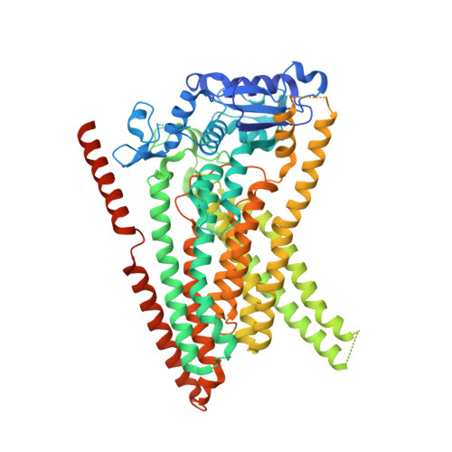
6R65

Crystal Structure of human TMEM16K / Anoctamin 10 (Form 2)


Experimental Data Snapshot

  • Method: X-RAY DIFFRACTION
  • Resolution: 3.50 Å
  • R-Value Free: 0.249 
  • R-Value Work: 0.237 
  • R-Value Observed: 0.238 

Starting Model: experimental
View more details

wwPDB Validation   3D Report Full Report


This is version 1.4 of the entry. See complete history


Literature

The structural basis of lipid scrambling and inactivation in the endoplasmic reticulum scramblase TMEM16K.

Bushell, S.R.Pike, A.C.W.Falzone, M.E.Rorsman, N.J.G.Ta, C.M.Corey, R.A.Newport, T.D.Christianson, J.C.Scofano, L.F.Shintre, C.A.Tessitore, A.Chu, A.Wang, Q.Shrestha, L.Mukhopadhyay, S.M.M.Love, J.D.Burgess-Brown, N.A.Sitsapesan, R.Stansfeld, P.J.Huiskonen, J.T.Tammaro, P.Accardi, A.Carpenter, E.P.

(2019) Nat Commun 10: 3956-3956

  • DOI: https://doi.org/10.1038/s41467-019-11753-1
  • Primary Citation of Related Structures:  
    5OC9, 6R65, 6R7X, 6R7Y, 6R7Z

  • PubMed Abstract: 

    Membranes in cells have defined distributions of lipids in each leaflet, controlled by lipid scramblases and flip/floppases. However, for some intracellular membranes such as the endoplasmic reticulum (ER) the scramblases have not been identified. Members of the TMEM16 family have either lipid scramblase or chloride channel activity. Although TMEM16K is widely distributed and associated with the neurological disorder autosomal recessive spinocerebellar ataxia type 10 (SCAR10), its location in cells, function and structure are largely uncharacterised. Here we show that TMEM16K is an ER-resident lipid scramblase with a requirement for short chain lipids and calcium for robust activity. Crystal structures of TMEM16K show a scramblase fold, with an open lipid transporting groove. Additional cryo-EM structures reveal extensive conformational changes from the cytoplasmic to the ER side of the membrane, giving a state with a closed lipid permeation pathway. Molecular dynamics simulations showed that the open-groove conformation is necessary for scramblase activity.


  • Organizational Affiliation

    Structural Genomics Consortium, Nuffield Department of Medicine, University of Oxford, Old Road Campus Research Building, Roosevelt Drive, Oxford, OX3 7DQ, UK.


Macromolecules
Find similar proteins by:  (by identity cutoff)  |  3D Structure
Entity ID: 1
MoleculeChains Sequence LengthOrganismDetailsImage
Anoctamin-10
A, B
667Homo sapiensMutation(s): 0 
Gene Names: ANO10TMEM16K
Membrane Entity: Yes 
UniProt & NIH Common Fund Data Resources
Find proteins for Q9NW15 (Homo sapiens)
Explore Q9NW15 
Go to UniProtKB:  Q9NW15
PHAROS:  Q9NW15
GTEx:  ENSG00000160746 
Entity Groups  
Sequence Clusters30% Identity50% Identity70% Identity90% Identity95% Identity100% Identity
UniProt GroupQ9NW15
Sequence Annotations
Expand
  • Reference Sequence
Experimental Data & Validation

Experimental Data

  • Method: X-RAY DIFFRACTION
  • Resolution: 3.50 Å
  • R-Value Free: 0.249 
  • R-Value Work: 0.237 
  • R-Value Observed: 0.238 
  • Space Group: P 21 21 21
Unit Cell:
Length ( Å )Angle ( ˚ )
a = 113.158α = 90
b = 152.267β = 90
c = 153.703γ = 90
Software Package:
Software NamePurpose
BUSTERrefinement
PDB_EXTRACTdata extraction
XDSdata reduction
XDSdata scaling
STARANISOdata scaling
PHASERphasing

Structure Validation

View Full Validation Report


Entry History & Funding Information

Deposition Data


Funding OrganizationLocationGrant Number
Wellcome TrustUnited Kingdom106169/Z/14/Z
European CommissionUnited Kingdom115766

Revision History  (Full details and data files)

  • Version 1.0: 2019-05-01
    Type: Initial release
  • Version 1.1: 2019-07-10
    Changes: Data collection
  • Version 1.2: 2019-10-16
    Changes: Data collection
  • Version 1.3: 2019-11-20
    Changes: Database references
  • Version 1.4: 2024-01-24
    Changes: Data collection, Database references, Derived calculations, Refinement description