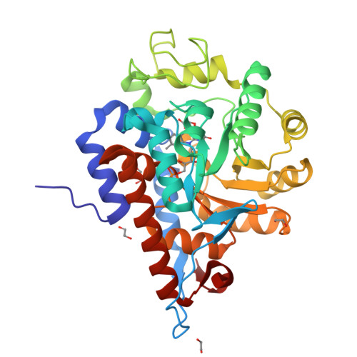
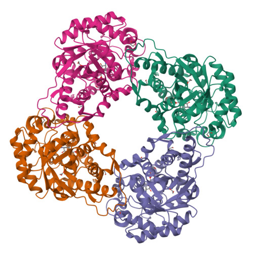
Structure of human hydroxyacid oxidase 1 bound with FMN and glycolate
MacKinnon, S., Bezerra, G.A., Krojer, T., Smee, C., Arrowsmith, C.H., Edwards, E., Bountra, C., Oppermann, U., Brennan, P.E., Yue, W.W.To be published.
Experimental Data Snapshot
Starting Model: experimental
View more details
Entity ID: 1 | |||||
|---|---|---|---|---|---|
| Molecule | Chains | Sequence Length | Organism | Details | Image |
| Hydroxyacid oxidase 1 | 362 | Homo sapiens | Mutation(s): 0 Gene Names: HAO1, GOX1, HAOX1 EC: 1.1.3.15 (PDB Primary Data), 1.2.3.5 (UniProt) | 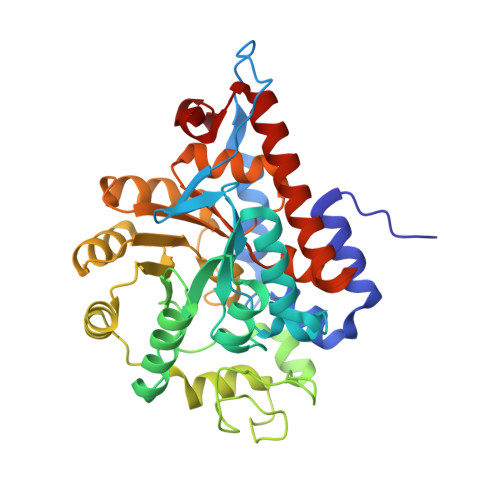 | |
UniProt & NIH Common Fund Data Resources | |||||
Find proteins for Q9UJM8 (Homo sapiens) Explore Q9UJM8 Go to UniProtKB: Q9UJM8 | |||||
PHAROS: Q9UJM8 GTEx: ENSG00000101323 | |||||
Entity Groups | |||||
| Sequence Clusters | 30% Identity50% Identity70% Identity90% Identity95% Identity100% Identity | ||||
| UniProt Group | Q9UJM8 | ||||
Sequence AnnotationsExpand | |||||
| |||||
| Ligands 4 Unique | |||||
|---|---|---|---|---|---|
| ID | Chains | Name / Formula / InChI Key | 2D Diagram | 3D Interactions | |
| FMN Query on FMN | G [auth A] | FLAVIN MONONUCLEOTIDE C17 H21 N4 O9 P FVTCRASFADXXNN-SCRDCRAPSA-N |  | ||
| PG4 Query on PG4 | D [auth A] | TETRAETHYLENE GLYCOL C8 H18 O5 UWHCKJMYHZGTIT-UHFFFAOYSA-N |  | ||
| GOA Query on GOA | H [auth A] | GLYCOLIC ACID C2 H4 O3 AEMRFAOFKBGASW-UHFFFAOYSA-N |  | ||
| EDO Query on EDO | B [auth A], C [auth A], E [auth A], F [auth A] | 1,2-ETHANEDIOL C2 H6 O2 LYCAIKOWRPUZTN-UHFFFAOYSA-N |  | ||
| Length ( Å ) | Angle ( ˚ ) |
|---|---|
| a = 97.431 | α = 90 |
| b = 97.431 | β = 90 |
| c = 80.364 | γ = 90 |
| Software Name | Purpose |
|---|---|
| REFMAC | refinement |
| SCALA | data scaling |
| PDB_EXTRACT | data extraction |
| SCALA | data scaling |
| PHASER | phasing |
| XDS | data reduction |
| Funding Organization | Location | Grant Number |
|---|---|---|
| Wellcome Trust | United Kingdom | 106169/ZZ14/Z |
RCSB PDB is hosted by

RCSB PDB is a member of the