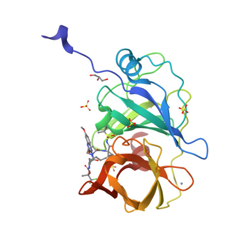
Design of Hepatitis C NS3/4A Protease Inhibitors Leveraging Untapped Regions of the Substrate Envelope
Matthew, A.N., Schiffer, C.A.To be published.
Experimental Data Snapshot
Starting Model: experimental
View more details
Entity ID: 1 | |||||
|---|---|---|---|---|---|
| Molecule | Chains | Sequence Length | Organism | Details | Image |
| NS3 protease | 199 | Hepacivirus hominis | Mutation(s): 0 | 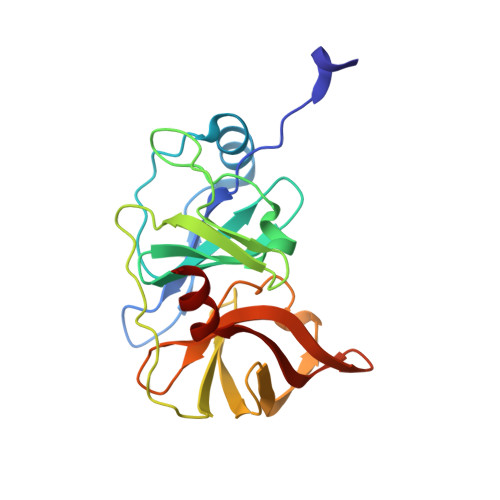 | |
UniProt | |||||
Find proteins for A0A0B4WYC6 (Hepacivirus hominis) Explore A0A0B4WYC6 Go to UniProtKB: A0A0B4WYC6 | |||||
Entity Groups | |||||
| Sequence Clusters | 30% Identity50% Identity70% Identity90% Identity95% Identity100% Identity | ||||
| UniProt Group | A0A0B4WYC6 | ||||
Sequence AnnotationsExpand | |||||
| |||||
| Ligands 4 Unique | |||||
|---|---|---|---|---|---|
| ID | Chains | Name / Formula / InChI Key | 2D Diagram | 3D Interactions | |
| GKG (Subject of Investigation/LOI) Query on GKG | B [auth A] | 1,1,1-trifluoro-2-methylpropan-2-yl
[(2R,6S,12Z,13aS,14aR,16aS)-2-[(7-methoxy-3-methylquinoxalin-2-yl)oxy]-14a-{[(1-methylcyclopropyl)sulfonyl]carbamoyl}-5,
16-dioxo-1,2,3,5,6,7,8,9,10,11,13a,14,14a,15,16,16a-hexadecahydrocyclopropa[e]pyrrolo[1,2-a][1,4]diazacyclopentadecin-6-
yl]carbamate C37 H47 F3 N6 O9 S ZOQBCVXDXFPSSL-DDAYHPHASA-N |  | ||
| SO4 Query on SO4 | C [auth A], D [auth A], E [auth A], F [auth A] | SULFATE ION O4 S QAOWNCQODCNURD-UHFFFAOYSA-L |  | ||
| GOL Query on GOL | H [auth A] | GLYCEROL C3 H8 O3 PEDCQBHIVMGVHV-UHFFFAOYSA-N |  | ||
| ZN Query on ZN | G [auth A] | ZINC ION Zn PTFCDOFLOPIGGS-UHFFFAOYSA-N |  | ||
| Length ( Å ) | Angle ( ˚ ) |
|---|---|
| a = 55.607 | α = 90 |
| b = 58.628 | β = 90 |
| c = 59.966 | γ = 90 |
| Software Name | Purpose |
|---|---|
| HKL-3000 | data scaling |
| PHASER | phasing |
| PHENIX | refinement |
| Coot | model building |
| Funding Organization | Location | Grant Number |
|---|---|---|
| National Institutes of Health/National Institute Of Allergy and Infectious Diseases (NIH/NIAID) | United States | R01 AI085051 |
| National Institutes of Health/National Institute of General Medical Sciences (NIH/NIGMS) | United States | F31 GM119345 |
RCSB PDB is hosted by

RCSB PDB is a member of the