6AAL | pdb_00006aal

Crystal Structure of putative amino acid binding periplasmic ABC transporter protein from Candidatus Liberibacter asiaticus in complex with Arginine


Experimental Data Snapshot

  • Method: X-RAY DIFFRACTION
  • Resolution: 2.60 Å
  • R-Value Free: 
    0.290 (Depositor), 0.290 (DCC) 
  • R-Value Work: 
    0.200 (Depositor), 0.200 (DCC) 
  • R-Value Observed: 
    0.205 (Depositor) 

Starting Model: experimental
View more details

wwPDB Validation   3D Report Full Report


Ligand Structure Quality Assessment 

Created with Raphaël 2.3.0Worse 01 BetterLigand structure goodness of fit to experimental dataBest fitted ARGClick on this verticalbar to view details

This is version 1.3 of the entry. See complete history


Literature

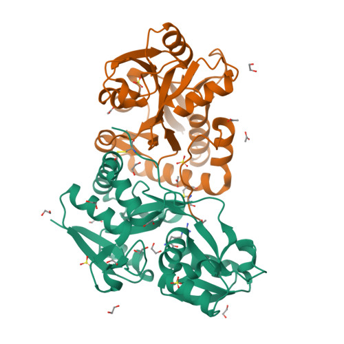
Crystal structures of a putative periplasmic cystine-binding protein from Candidatus Liberibacter asiaticus: insights into an adapted mechanism of ligand binding.

Kumar, P.Kesari, P.Kokane, S.Ghosh, D.K.Kumar, P.Sharma, A.K.

(2019) FEBS J 286: 3450-3472

  • DOI: https://doi.org/10.1111/febs.14921
  • Primary Citation of Related Structures:  
    6A80, 6A8S, 6AA1, 6AAL

  • PubMed Abstract: 

    The amino acid-binding receptors, a component of ABC transporters, have evolved to cater to different specificities and functions. Of particular interest are cystine-binding receptors, which have shown broad specificity. In the present study, a putative periplasmic cystine-binding protein from Candidatus Liberibacter asiaticus (CLasTcyA) was characterized. Analysis of the CLasTcyA sequence and crystal structures in the ligand-bound state revealed novel features of CLasTcyA in comparison to related proteins. One of the unique features found in CLasTcyA structure was the positioning of the C-terminal extended loop of one chain very close to the substrate-binding site of the adjacent monomer in the asymmetric unit. The presence of a disulphide bond, unique to Candidatus Liberibacter family, holds the C-terminal extended loop in position. Analysis of the substrate-binding pocket of CLasTcyA suggested a broad specificity and a completely different orientation of the bound substrates in comparison to related protein structures. The open conformation for one of the two chains of the asymmetric unit in the Arg-bound structure revealed a limited open state (18.4°) for CLasTcyA as compared to open state of other related proteins (~ 60°). The strong interaction between Asp126 on helix-α5 of small domain and Arg82 (bigger domain) restricts the degree of opening in ligand-free open state. The dissociation constant of 1.26 μm by SPR and 3.7 μm by MST exhibited low affinity for the cystine. This is the first structural characterization of an l-cystine ABC transporter from plant pathogen and our results suggest that CLasTcyA may have evolved to cater to its specific needs for its survival in the host.


  • Organizational Affiliation

    Department of Biotechnology, Indian Institute of Technology Roorkee, India.


Macromolecules
Find similar proteins by:  (by identity cutoff)  |  3D Structure
Entity ID: 1
MoleculeChains Sequence LengthOrganismDetailsImage
Putative amino acid-binding periplasmic ABC transporter protein
A, B
241Candidatus Liberibacter asiaticus str. psy62Mutation(s): 0 
Gene Names: CLIBASIA_05070
UniProt
Find proteins for C6XGT2 (Liberibacter asiaticus (strain psy62))
Explore C6XGT2 
Go to UniProtKB:  C6XGT2
Entity Groups  
Sequence Clusters30% Identity50% Identity70% Identity90% Identity95% Identity100% Identity
UniProt GroupC6XGT2
Sequence Annotations
Expand
  • Reference Sequence
Small Molecules
Ligands 4 Unique
IDChains Name / Formula / InChI Key2D Diagram3D Interactions
ARG
Query on ARG

Download Ideal Coordinates CCD File 
F [auth A]ARGININE
C6 H15 N4 O2
ODKSFYDXXFIFQN-BYPYZUCNSA-O
SO4
Query on SO4

Download Ideal Coordinates CCD File 
D [auth A],
E [auth A],
Q [auth B],
R [auth B]
SULFATE ION
O4 S
QAOWNCQODCNURD-UHFFFAOYSA-L
EDO
Query on EDO

Download Ideal Coordinates CCD File 
C [auth A]
G [auth A]
H [auth A]
I [auth A]
J [auth A]
1,2-ETHANEDIOL
C2 H6 O2
LYCAIKOWRPUZTN-UHFFFAOYSA-N
ACT
Query on ACT

Download Ideal Coordinates CCD File 
P [auth A],
T [auth B],
U [auth B],
V [auth B],
W [auth B]
ACETATE ION
C2 H3 O2
QTBSBXVTEAMEQO-UHFFFAOYSA-M
Experimental Data & Validation

Experimental Data

  • Method: X-RAY DIFFRACTION
  • Resolution: 2.60 Å
  • R-Value Free:  0.290 (Depositor), 0.290 (DCC) 
  • R-Value Work:  0.200 (Depositor), 0.200 (DCC) 
  • R-Value Observed: 0.205 (Depositor) 
Space Group: P 21 21 21
Unit Cell:
Length ( Å )Angle ( ˚ )
a = 46.342α = 90
b = 86β = 90
c = 123.832γ = 90
Software Package:
Software NamePurpose
REFMACrefinement
HKL-2000data reduction
HKL-2000data scaling
MoRDaphasing

Structure Validation

View Full Validation Report


Ligand Structure Quality Assessment 

Created with Raphaël 2.3.0Worse 01 BetterLigand structure goodness of fit to experimental dataBest fitted ARGClick on this verticalbar to view details

Entry History 

Deposition Data

Revision History  (Full details and data files)

  • Version 1.0: 2019-06-12
    Type: Initial release
  • Version 1.1: 2019-09-18
    Changes: Data collection, Database references
  • Version 1.2: 2023-11-22
    Changes: Data collection, Database references, Refinement description
  • Version 1.3: 2024-11-20
    Changes: Structure summary