5TP9

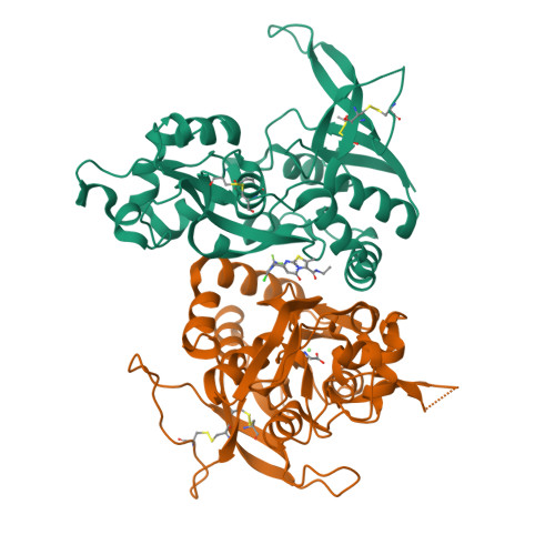
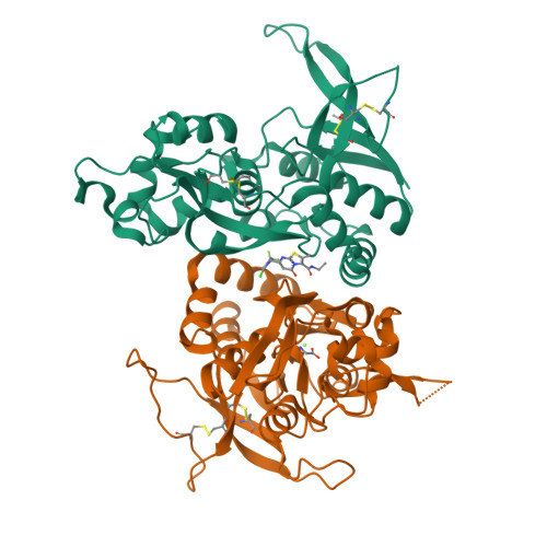
Structure of the human GluN1/GluN2A LBD in complex with compound 2 (GNE9178)


Experimental Data Snapshot

  • Method: X-RAY DIFFRACTION
  • Resolution: 2.40 Å
  • R-Value Free: 0.237 
  • R-Value Work: 0.190 
  • R-Value Observed: 0.192 

wwPDB Validation   3D Report Full Report


Ligand Structure Quality Assessment 


This is version 1.3 of the entry. See complete history


Literature

GluN2A-Selective Pyridopyrimidinone Series of NMDAR Positive Allosteric Modulators with an Improved in Vivo Profile.

Villemure, E.Volgraf, M.Jiang, Y.Wu, G.Ly, C.Q.Yuen, P.W.Lu, A.Luo, X.Liu, M.Zhang, S.Lupardus, P.J.Wallweber, H.J.Liederer, B.M.Deshmukh, G.Plise, E.Tay, S.Wang, T.M.Hanson, J.E.Hackos, D.H.Scearce-Levie, K.Schwarz, J.B.Sellers, B.D.

(2017) ACS Med Chem Lett 8: 84-89

  • DOI: https://doi.org/10.1021/acsmedchemlett.6b00388
  • Primary Citation of Related Structures:  
    5TP9, 5TPA

  • PubMed Abstract: 

    The N -methyl-d-aspartate receptor (NMDAR) is an ionotropic glutamate receptor, gated by the endogenous coagonists glutamate and glycine, permeable to Ca 2+ and Na + . NMDAR dysfunction is associated with numerous neurological and psychiatric disorders, including schizophrenia, depression, and Alzheimer's disease. Recently, we have disclosed GNE-0723 ( 1 ), a GluN2A subunit-selective and brain-penetrant positive allosteric modulator (PAM) of NMDARs. This work highlights the discovery of a related pyridopyrimidinone core with distinct structure-activity relationships, despite the structural similarity to GNE-0723. GNE-5729 ( 13 ), a pyridopyrimidinone-based NMDAR PAM, was identified with both an improved pharmacokinetic profile and increased selectivity against AMPARs. We also include X-ray structure analysis and modeling to propose hypotheses for the activity and selectivity differences.


  • Organizational Affiliation

    Department of Discovery Chemistry, Department of Neurosciences, Department of Biochemical and Cellular Pharmacology, Department of Drug Metabolism and Pharmacokinetics, and Department of Structural Biology, Genentech, Inc. , 1 DNA Way, South San Francisco, California 94080, United States.


Macromolecules
Find similar proteins by:  (by identity cutoff)  |  3D Structure
Entity ID: 1
MoleculeChains Sequence LengthOrganismDetailsImage
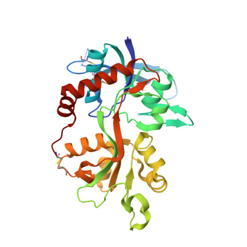
Glutamate receptor ionotropic, NMDA 2A285Homo sapiensMutation(s): 0 
Gene Names: GRIN2ANMDAR2A
UniProt & NIH Common Fund Data Resources
Find proteins for Q12879 (Homo sapiens)
Explore Q12879 
Go to UniProtKB:  Q12879
PHAROS:  Q12879
GTEx:  ENSG00000183454 
Entity Groups  
Sequence Clusters30% Identity50% Identity70% Identity90% Identity95% Identity100% Identity
UniProt GroupQ12879
Sequence Annotations
Expand
  • Reference Sequence
Find similar proteins by:  (by identity cutoff)  |  3D Structure
Entity ID: 2
MoleculeChains Sequence LengthOrganismDetailsImage
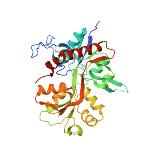
Glutamate receptor ionotropic, NMDA 1293Homo sapiensMutation(s): 0 
Gene Names: GRIN1NMDAR1
UniProt & NIH Common Fund Data Resources
Find proteins for Q05586 (Homo sapiens)
Explore Q05586 
Go to UniProtKB:  Q05586
PHAROS:  Q05586
GTEx:  ENSG00000176884 
Entity Groups  
Sequence Clusters30% Identity50% Identity70% Identity90% Identity95% Identity100% Identity
UniProt GroupQ05586
Sequence Annotations
Expand
  • Reference Sequence
Small Molecules
Ligands 5 Unique
IDChains Name / Formula / InChI Key2D Diagram3D Interactions
7H0
Query on 7H0

Download Ideal Coordinates CCD File 
E [auth A]7-{[5-chloro-3-(trifluoromethyl)-1H-pyrazol-1-yl]methyl}-N-ethyl-2-methyl-5-oxo-5H-[1,3]thiazolo[3,2-a]pyrimidine-3-carboxamide
C15 H13 Cl F3 N5 O2 S
MQDBLHZAPZLITE-UHFFFAOYSA-N
GLU
Query on GLU

Download Ideal Coordinates CCD File 
D [auth A]GLUTAMIC ACID
C5 H9 N O4
WHUUTDBJXJRKMK-VKHMYHEASA-N
GLY
Query on GLY

Download Ideal Coordinates CCD File 
G [auth B]GLYCINE
C2 H5 N O2
DHMQDGOQFOQNFH-UHFFFAOYSA-N
ACT
Query on ACT

Download Ideal Coordinates CCD File 
C [auth A]ACETATE ION
C2 H3 O2
QTBSBXVTEAMEQO-UHFFFAOYSA-M
CA
Query on CA

Download Ideal Coordinates CCD File 
F [auth B]CALCIUM ION
Ca
BHPQYMZQTOCNFJ-UHFFFAOYSA-N
Binding Affinity Annotations 
IDSourceBinding Affinity
7H0 BindingDB:  5TP9 EC50: min: 436, max: 440 (nM) from 2 assay(s)
Experimental Data & Validation

Experimental Data

  • Method: X-RAY DIFFRACTION
  • Resolution: 2.40 Å
  • R-Value Free: 0.237 
  • R-Value Work: 0.190 
  • R-Value Observed: 0.192 
  • Space Group: P 21 21 21
Unit Cell:
Length ( Å )Angle ( ˚ )
a = 56.26α = 90
b = 89.78β = 90
c = 120.86γ = 90
Software Package:
Software NamePurpose
BUSTERrefinement
HKL-2000data reduction
SCALEPACKdata scaling
PHASERphasing

Structure Validation

View Full Validation Report


Ligand Structure Quality Assessment 


Entry History 

Deposition Data

Revision History  (Full details and data files)

  • Version 1.0: 2016-11-30
    Type: Initial release
  • Version 1.1: 2016-12-28
    Changes: Database references
  • Version 1.2: 2017-02-08
    Changes: Database references
  • Version 1.3: 2024-10-16
    Changes: Data collection, Database references, Structure summary