5ZW8

PigA with FAD and proline


Experimental Data Snapshot

  • Method: X-RAY DIFFRACTION
  • Resolution: 1.69 Å
  • R-Value Free: 
    0.180 (Depositor), 0.180 (DCC) 
  • R-Value Work: 
    0.154 (Depositor), 0.150 (DCC) 
  • R-Value Observed: 
    0.155 (Depositor) 

Starting Model: experimental
View more details

wwPDB Validation   3D Report Full Report


Ligand Structure Quality Assessment 

Created with Raphaël 2.3.0Worse 01 BetterLigand structure goodness of fit to experimental dataBest fitted FADClick on this verticalbar to view detailsBest fitted O4BClick on this verticalbar to view details

This is version 1.2 of the entry. See complete history


Literature

Crystal Structure of PigA: A Prolyl Thioester-Oxidizing Enzyme in Prodigiosin Biosynthesis.

Lee, C.-C.Ko, T.-P.Chen, C.T.Chan, Y.T.Lo, S.Y.Chang, J.Y.Chen, Y.W.Chung, T.F.Hsieh, H.J.Hsiao, C.D.Wang, A.H.J.

(2019) Chembiochem 20: 193-202

  • DOI: https://doi.org/10.1002/cbic.201800409
  • Primary Citation of Related Structures:  
    5ZW0, 5ZW2, 5ZW7, 5ZW8, 6AF6

  • PubMed Abstract: 

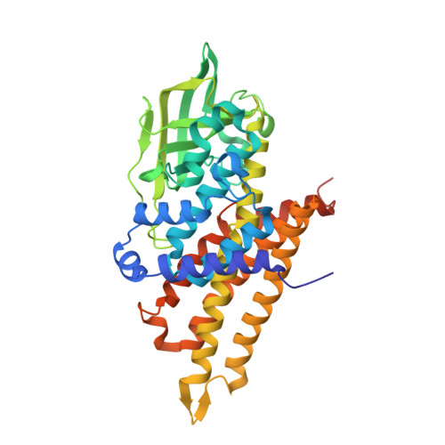
    Prodigiosin is an intensely red pigment comprising three pyrroles. The biosynthetic pathway includes a two-step proline oxidation catalyzed by phosphatidylinositol N-acetylglucosaminyltransferase subunit A (PigA), with flavin adenine dinucleotide (FAD) as its cofactor. The enzyme is crystallized in the apo form and in complex with FAD and proline. As an acyl coenzyme A dehydrogenase (ACAD) family member, the protein folds into a β-sheet flanked by two α-helical domains. PigA forms a tetramer, which is consistent with analytical ultracentrifugation results. FAD binds to PigA in a similar way to that in the other enzymes of the ACAD family. The variable conformations of loop β4-β5 and helix αG correlate well with the structural flexibility required for substrate entrance to the Re side of FAD. Modeling with PigG, the acyl carrier protein, suggests a reasonable mode of interaction with PigA. The structure helps to explain the proline oxidation mechanism, in which Glu244 plays a central role by abstracting the substrate protons. It also reveals a plausible pocket for oxygen binding to the Si side of FAD.


  • Organizational Affiliation

    Institute of Biological Chemistry, Academia Sinica, 128 Academia Road Section 2, Taipei, 11529, Taiwan.


Macromolecules
Find similar proteins by:  (by identity cutoff)  |  3D Structure
Entity ID: 1
MoleculeChains Sequence LengthOrganismDetailsImage
L-prolyl-[peptidyl-carrier protein] dehydrogenase402Prodigiosinella confusarubidaMutation(s): 0 
Gene Names: pigA
EC: 1.3.8.14
UniProt
Find proteins for Q5W271 (Serratia sp. (strain ATCC 39006))
Explore Q5W271 
Go to UniProtKB:  Q5W271
Entity Groups  
Sequence Clusters30% Identity50% Identity70% Identity90% Identity95% Identity100% Identity
UniProt GroupQ5W271
Sequence Annotations
Expand
  • Reference Sequence
Small Molecules
Ligands 5 Unique
IDChains Name / Formula / InChI Key2D Diagram3D Interactions
FAD
Query on FAD

Download Ideal Coordinates CCD File 
B [auth A]FLAVIN-ADENINE DINUCLEOTIDE
C27 H33 N9 O15 P2
VWWQXMAJTJZDQX-UYBVJOGSSA-N
O4B
Query on O4B

Download Ideal Coordinates CCD File 
C [auth A],
D [auth A]
1,4,7,10,13,16-HEXAOXACYCLOOCTADECANE
C12 H24 O6
XEZNGIUYQVAUSS-UHFFFAOYSA-N
PRO
Query on PRO

Download Ideal Coordinates CCD File 
G [auth A]PROLINE
C5 H9 N O2
ONIBWKKTOPOVIA-BYPYZUCNSA-N
MLA
Query on MLA

Download Ideal Coordinates CCD File 
E [auth A]MALONIC ACID
C3 H4 O4
OFOBLEOULBTSOW-UHFFFAOYSA-N
CL
Query on CL

Download Ideal Coordinates CCD File 
F [auth A]CHLORIDE ION
Cl
VEXZGXHMUGYJMC-UHFFFAOYSA-M
Experimental Data & Validation

Experimental Data

  • Method: X-RAY DIFFRACTION
  • Resolution: 1.69 Å
  • R-Value Free:  0.180 (Depositor), 0.180 (DCC) 
  • R-Value Work:  0.154 (Depositor), 0.150 (DCC) 
  • R-Value Observed: 0.155 (Depositor) 
Space Group: I 2 2 2
Unit Cell:
Length ( Å )Angle ( ˚ )
a = 85.585α = 90
b = 85.558β = 90
c = 146.144γ = 90
Software Package:
Software NamePurpose
PHENIXrefinement
HKL-2000data reduction
HKL-2000data scaling
PHENIXphasing

Structure Validation

View Full Validation Report


Ligand Structure Quality Assessment 

Created with Raphaël 2.3.0Worse 01 BetterLigand structure goodness of fit to experimental dataBest fitted FADClick on this verticalbar to view detailsBest fitted O4BClick on this verticalbar to view details

Entry History & Funding Information

Deposition Data


Funding OrganizationLocationGrant Number
Taiwan--

Revision History  (Full details and data files)

  • Version 1.0: 2018-09-05
    Type: Initial release
  • Version 1.1: 2019-02-06
    Changes: Data collection, Database references
  • Version 1.2: 2023-11-22
    Changes: Data collection, Database references, Refinement description