


X-ray snapshots of two classes of cysteine desulfurase enzymes NifS and SufS
Nakamura, R., Fujishiro, T., Takahashi, Y.To be published.
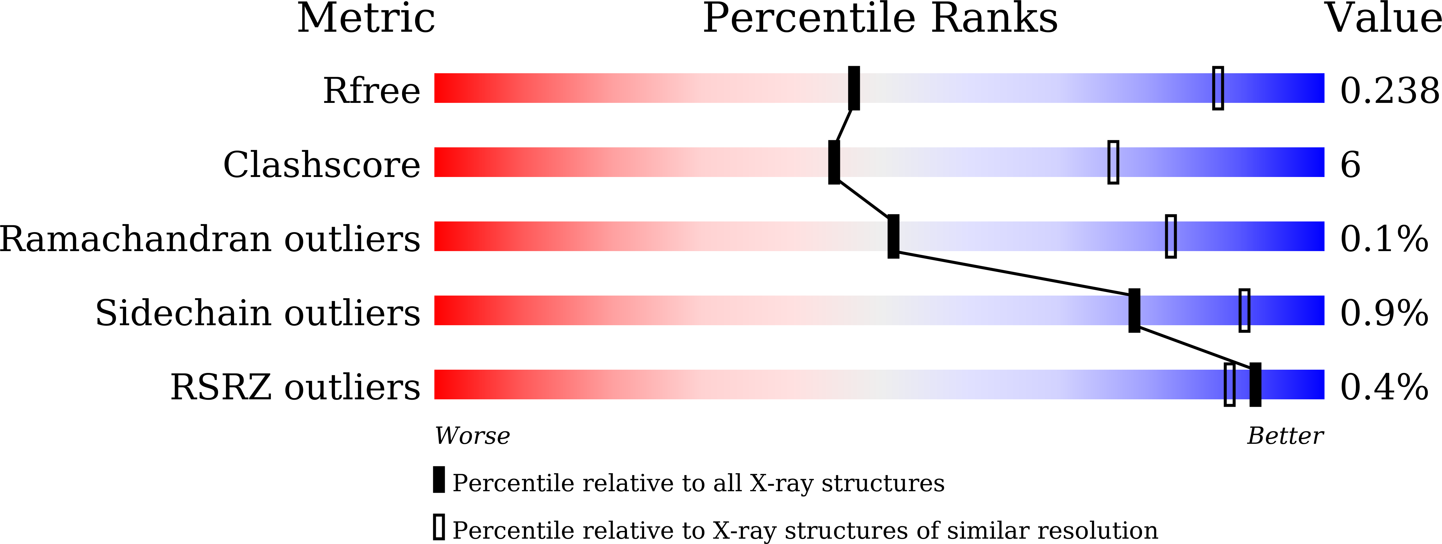
Experimental Data Snapshot
Starting Model: experimental
View more details
Entity ID: 1 | |||||
|---|---|---|---|---|---|
| Molecule | Chains | Sequence Length | Organism | Details | Image |
| Cysteine desulfurase | 415 | Hydrogenimonas thermophila | Mutation(s): 0 Gene Names: SAMN05216234_11013 EC: 2.8.1.7 | 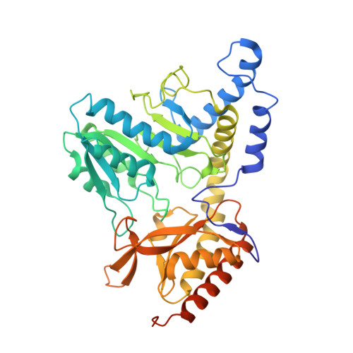 | |
UniProt | |||||
Find proteins for A0A1I5NEH3 (Hydrogenimonas thermophila) Explore A0A1I5NEH3 Go to UniProtKB: A0A1I5NEH3 | |||||
Entity Groups | |||||
| Sequence Clusters | 30% Identity50% Identity70% Identity90% Identity95% Identity100% Identity | ||||
| UniProt Group | A0A1I5NEH3 | ||||
Sequence AnnotationsExpand | |||||
| |||||
Entity ID: 2 | |||||
|---|---|---|---|---|---|
| Molecule | Chains | Sequence Length | Organism | Details | Image |
| Cysteine desulfurase | 415 | Hydrogenimonas thermophila | Mutation(s): 0 Gene Names: SAMN05216234_11013 EC: 2.8.1.7 | 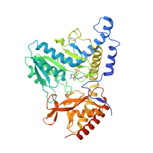 | |
UniProt | |||||
Find proteins for A0A1I5NEH3 (Hydrogenimonas thermophila) Explore A0A1I5NEH3 Go to UniProtKB: A0A1I5NEH3 | |||||
Entity Groups | |||||
| Sequence Clusters | 30% Identity50% Identity70% Identity90% Identity95% Identity100% Identity | ||||
| UniProt Group | A0A1I5NEH3 | ||||
Sequence AnnotationsExpand | |||||
| |||||
| Ligands 1 Unique | |||||
|---|---|---|---|---|---|
| ID | Chains | Name / Formula / InChI Key | 2D Diagram | 3D Interactions | |
| C6P Query on C6P | C [auth A] | N-({3-HYDROXY-2-METHYL-5-[(PHOSPHONOOXY)METHYL]PYRIDIN-4-YL}METHYL)-L-CYSTEINE C11 H17 N2 O7 P S FPVGQJHHLSVHOT-VIFPVBQESA-N |  | ||
| Modified Residues 1 Unique | |||||
|---|---|---|---|---|---|
| ID | Chains | Type | Formula | 2D Diagram | Parent |
| LLP Query on LLP | B | L-PEPTIDE LINKING | C14 H22 N3 O7 P |  | LYS |
| Length ( Å ) | Angle ( ˚ ) |
|---|---|
| a = 138.2 | α = 90 |
| b = 138.2 | β = 90 |
| c = 99.1 | γ = 120 |
| Software Name | Purpose |
|---|---|
| PHENIX | refinement |
| XSCALE | data scaling |
| PDB_EXTRACT | data extraction |
| XDS | data reduction |
| MOLREP | phasing |
| Funding Organization | Location | Grant Number |
|---|---|---|
| Japan Society for the Promotion of Science | Japan | 17K14510 |
| Japan Society for the Promotion of Science | Japan | 15H04472 |
RCSB PDB is hosted by

RCSB PDB is a member of the


