5MJH

X-ray generated oxyferrous/water mixed complex of DtpA from Streptomyces lividans


Experimental Data Snapshot

  • Method: X-RAY DIFFRACTION
  • Resolution: 1.45 Å
  • R-Value Free: 
    0.254 (Depositor), 0.260 (DCC) 
  • R-Value Work: 
    0.221 (Depositor), 0.230 (DCC) 
  • R-Value Observed: 
    0.222 (Depositor) 

wwPDB Validation   3D Report Full Report


Ligand Structure Quality Assessment 

Created with Raphaël 2.3.0Worse 01 BetterLigand structure goodness of fit to experimental dataBest fitted HEMClick on this verticalbar to view details

This is version 1.2 of the entry. See complete history


Literature

Photoreduction and validation of haem-ligand intermediate states in protein crystals by in situ single-crystal spectroscopy and diffraction.

Kekilli, D.Moreno-Chicano, T.Chaplin, A.K.Horrell, S.Dworkowski, F.S.N.Worrall, J.A.R.Strange, R.W.Hough, M.A.

(2017) IUCrJ 4: 263-270

  • DOI: https://doi.org/10.1107/S2052252517002159
  • Primary Citation of Related Structures:  
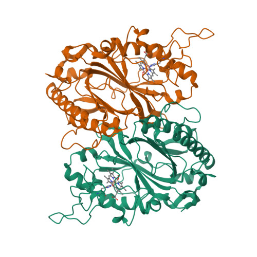
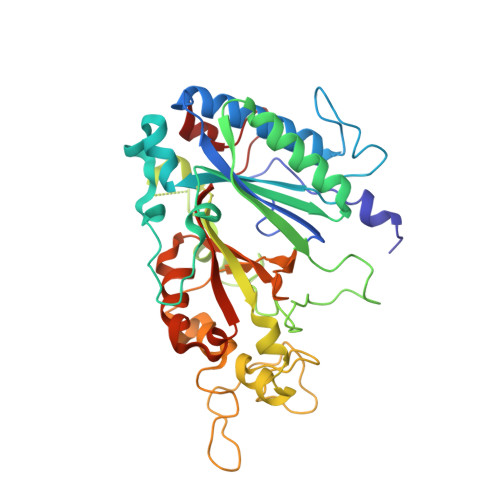
    5MAP, 5MJH

  • PubMed Abstract: 

    Powerful synergies are available from the combination of multiple methods to study proteins in the crystalline form. Spectroscopies which probe the same region of the crystal from which X-ray crystal structures are determined can give insights into redox, ligand and spin states to complement the information gained from the electron-density maps. The correct assignment of crystal structures to the correct protein redox and ligand states is essential to avoid the misinterpretation of structural data. This is a particular concern for haem proteins, which can occupy a wide range of redox states and are exquisitely sensitive to becoming reduced by solvated electrons generated from interactions of X-rays with water molecules in the crystal. Here, single-crystal spectroscopic fingerprinting has been applied to investigate the laser photoreduction of ferric haem in cytochrome c '. Furthermore, in situ X-ray-driven generation of haem intermediates in crystals of the dye-decolourizing-type peroxidase A (DtpA) from Streptomyces lividans is described.


  • Organizational Affiliation

    School of Biological Sciences, University of Essex, Wivenhoe Park, Colchester CO4 3SQ, England.


Macromolecules
Find similar proteins by:  (by identity cutoff)  |  3D Structure
Entity ID: 1
MoleculeChains Sequence LengthOrganismDetailsImage
Dye type peroxidase A
A, B
377Streptomyces lividans 1326Mutation(s): 0 
Gene Names: SLIV_18505
Entity Groups  
Sequence Clusters30% Identity50% Identity70% Identity90% Identity95% Identity100% Identity
Sequence Annotations
Expand
  • Reference Sequence
Experimental Data & Validation

Experimental Data

  • Method: X-RAY DIFFRACTION
  • Resolution: 1.45 Å
  • R-Value Free:  0.254 (Depositor), 0.260 (DCC) 
  • R-Value Work:  0.221 (Depositor), 0.230 (DCC) 
  • R-Value Observed: 0.222 (Depositor) 
Space Group: P 1 21 1
Unit Cell:
Length ( Å )Angle ( ˚ )
a = 59.782α = 90
b = 70.627β = 93.01
c = 77.642γ = 90
Software Package:
Software NamePurpose
REFMACrefinement
Aimlessdata scaling
PDB_EXTRACTdata extraction
XDSdata reduction
MOLREPphasing

Structure Validation

View Full Validation Report


Ligand Structure Quality Assessment 

Created with Raphaël 2.3.0Worse 01 BetterLigand structure goodness of fit to experimental dataBest fitted HEMClick on this verticalbar to view details

Entry History 

Deposition Data

Revision History  (Full details and data files)

  • Version 1.0: 2017-05-10
    Type: Initial release
  • Version 1.1: 2017-05-31
    Changes: Database references
  • Version 1.2: 2024-05-08
    Changes: Advisory, Data collection, Database references, Derived calculations