5HR4 | pdb_00005hr4

Structure of Type IIL restriction-modification enzyme MmeI in complex with DNA has implications for engineering of new specificities


Experimental Data Snapshot

  • Method: X-RAY DIFFRACTION
  • Resolution: 2.60 Å
  • R-Value Free: 
    0.242 (Depositor), 0.240 (DCC) 
  • R-Value Work: 
    0.215 (Depositor), 0.220 (DCC) 
  • R-Value Observed: 
    0.216 (Depositor) 

wwPDB Validation   3D Report Full Report


Ligand Structure Quality Assessment 

Created with Raphaël 2.3.0Worse 01 BetterLigand structure goodness of fit to experimental dataBest fitted SFGClick on this verticalbar to view details

This is version 1.1 of the entry. See complete history


Literature

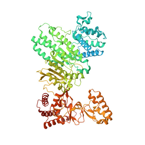
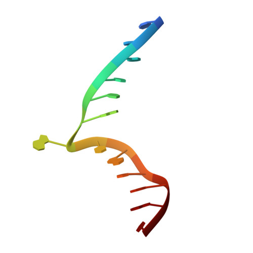
Structure of Type IIL Restriction-Modification Enzyme MmeI in Complex with DNA Has Implications for Engineering New Specificities.

Callahan, S.J.Luyten, Y.A.Gupta, Y.K.Wilson, G.G.Roberts, R.J.Morgan, R.D.Aggarwal, A.K.

(2016) PLoS Biol 14: e1002442-e1002442

  • DOI: https://doi.org/10.1371/journal.pbio.1002442
  • Primary Citation of Related Structures:  
    5HR4

  • PubMed Abstract: 

    The creation of restriction enzymes with programmable DNA-binding and -cleavage specificities has long been a goal of modern biology. The recently discovered Type IIL MmeI family of restriction-and-modification (RM) enzymes that possess a shared target recognition domain provides a framework for engineering such new specificities. However, a lack of structural information on Type IIL enzymes has limited the repertoire that can be rationally engineered. We report here a crystal structure of MmeI in complex with its DNA substrate and an S-adenosylmethionine analog (Sinefungin). The structure uncovers for the first time the interactions that underlie MmeI-DNA recognition and methylation (5'-TCCRAC-3'; R = purine) and provides a molecular basis for changing specificity at four of the six base pairs of the recognition sequence (5'-TCCRAC-3'). Surprisingly, the enzyme is resilient to specificity changes at the first position of the recognition sequence (5'-TCCRAC-3'). Collectively, the structure provides a basis for engineering further derivatives of MmeI and delineates which base pairs of the recognition sequence are more amenable to alterations than others.


  • Organizational Affiliation

    Department of Structural and Chemical Biology, Mount Sinai School of Medicine, New York, New York, United States of America.


Macromolecules

Find similar proteins by:  (by identity cutoff)  |  3D Structure
Entity ID: 1
MoleculeChains Sequence LengthOrganismDetailsImage
MmeIA [auth C],
D [auth J]
919Methylophilus methylotrophusMutation(s): 0 
Gene Names: mmeIRM
EC: 2.1.1.72
UniProt
Find proteins for B2MU09 (Methylophilus methylotrophus)
Explore B2MU09 
Go to UniProtKB:  B2MU09
Entity Groups  
Sequence Clusters30% Identity50% Identity70% Identity90% Identity95% Identity100% Identity
UniProt GroupB2MU09
Sequence Annotations
Expand
  • Reference Sequence
Experimental Data & Validation

Experimental Data

  • Method: X-RAY DIFFRACTION
  • Resolution: 2.60 Å
  • R-Value Free:  0.242 (Depositor), 0.240 (DCC) 
  • R-Value Work:  0.215 (Depositor), 0.220 (DCC) 
  • R-Value Observed: 0.216 (Depositor) 
Space Group: P 1
Unit Cell:
Length ( Å )Angle ( ˚ )
a = 61.877α = 72.84
b = 95.292β = 89.15
c = 161.969γ = 71.62
Software Package:
Software NamePurpose
PHENIXrefinement
XDSdata reduction
XDSdata scaling
PHENIXphasing

Structure Validation

View Full Validation Report


Ligand Structure Quality Assessment 

Created with Raphaël 2.3.0Worse 01 BetterLigand structure goodness of fit to experimental dataBest fitted SFGClick on this verticalbar to view details

Entry History 

Deposition Data

Revision History  (Full details and data files)

  • Version 1.0: 2016-04-27
    Type: Initial release
  • Version 1.1: 2024-03-06
    Changes: Data collection, Database references, Derived calculations