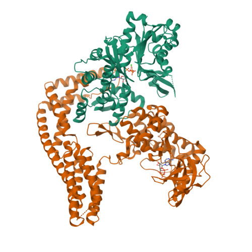
The Yersinia YopO - actin complex with MgADP
Lee, W.L., Grimes, J.M., Robinson, R.C.To be published.
Experimental Data Snapshot
Starting Model: experimental
View more details
Entity ID: 1 | |||||
|---|---|---|---|---|---|
| Molecule | Chains | Sequence Length | Organism | Details | Image |
| Actin | 376 | Spodoptera frugiperda | Mutation(s): 0 | 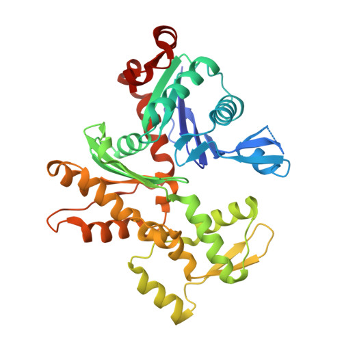 | |
UniProt | |||||
Find proteins for G3CKA6 (Spodoptera frugiperda) Explore G3CKA6 Go to UniProtKB: G3CKA6 | |||||
Entity Groups | |||||
| Sequence Clusters | 30% Identity50% Identity70% Identity90% Identity95% Identity100% Identity | ||||
| UniProt Group | G3CKA6 | ||||
Sequence AnnotationsExpand | |||||
| |||||
Entity ID: 2 | |||||
|---|---|---|---|---|---|
| Molecule | Chains | Sequence Length | Organism | Details | Image |
| Protein kinase YopO | 643 | Yersinia enterocolitica | Mutation(s): 5 Gene Names: yopO | 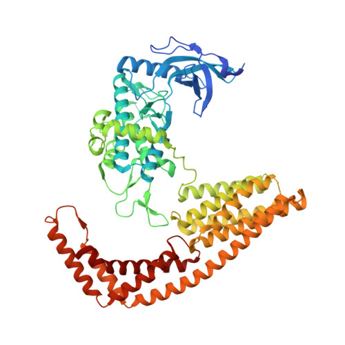 | |
UniProt | |||||
Find proteins for Q93KQ6 (Yersinia enterocolitica) Explore Q93KQ6 Go to UniProtKB: Q93KQ6 | |||||
Entity Groups | |||||
| Sequence Clusters | 30% Identity50% Identity70% Identity90% Identity95% Identity100% Identity | ||||
| UniProt Group | Q93KQ6 | ||||
Sequence AnnotationsExpand | |||||
| |||||
| Ligands 4 Unique | |||||
|---|---|---|---|---|---|
| ID | Chains | Name / Formula / InChI Key | 2D Diagram | 3D Interactions | |
| ATP Query on ATP | E [auth A], I [auth C] | ADENOSINE-5'-TRIPHOSPHATE C10 H16 N5 O13 P3 ZKHQWZAMYRWXGA-KQYNXXCUSA-N |  | ||
| ADP Query on ADP | G [auth B], K [auth D] | ADENOSINE-5'-DIPHOSPHATE C10 H15 N5 O10 P2 XTWYTFMLZFPYCI-KQYNXXCUSA-N |  | ||
| CA Query on CA | F [auth A], J [auth C] | CALCIUM ION Ca BHPQYMZQTOCNFJ-UHFFFAOYSA-N |  | ||
| MG Query on MG | H [auth B], L [auth D] | MAGNESIUM ION Mg JLVVSXFLKOJNIY-UHFFFAOYSA-N |  | ||
| Length ( Å ) | Angle ( ˚ ) |
|---|---|
| a = 108.73 | α = 90 |
| b = 121.75 | β = 104.88 |
| c = 118.58 | γ = 90 |
| Software Name | Purpose |
|---|---|
| PHENIX | refinement |
| xia2 | data reduction |
| xia2 | data scaling |
| PHASER | phasing |
| REFMAC | refinement |
| Funding Organization | Location | Grant Number |
|---|---|---|
| Agency for Science, Technology and Research (A*STAR) | Singapore | -- |
| Wellcome Trust | United Kingdom | 090532/Z/09/Z |