4YWY

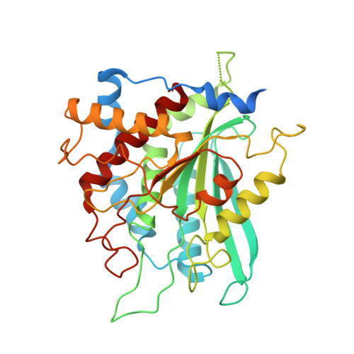
Crystal Structure of double mutant Y115E Y117E human Glutaminyl Cyclase in complex with inhibitor PBD-150


Experimental Data Snapshot

  • Method: X-RAY DIFFRACTION
  • Resolution: 1.95 Å
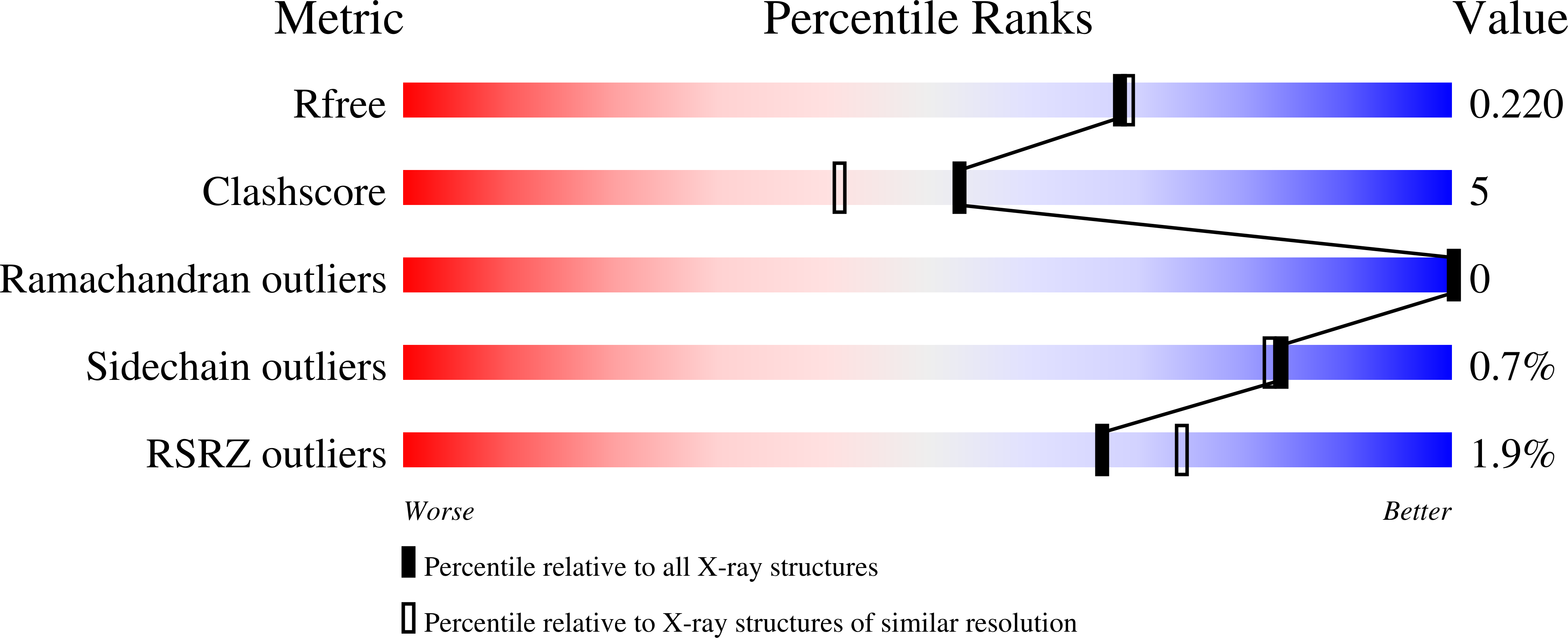
  • R-Value Free: 0.211 
  • R-Value Work: 0.164 
  • R-Value Observed: 0.166 

Starting Model: experimental
View more details

wwPDB Validation   3D Report Full Report


Ligand Structure Quality Assessment 


This is version 1.3 of the entry. See complete history


Literature

The soluble Y115E-Y117E variant of human glutaminyl cyclase is a valid target for X-ray and NMR screening of inhibitors against Alzheimer disease.

DiPisa, F.Pozzi, C.Benvenuti, M.Andreini, M.Marconi, G.Mangani, S.

(2015) Acta Crystallogr F Struct Biol Commun 71: 986-992

  • DOI: https://doi.org/10.1107/S2053230X15010389
  • Primary Citation of Related Structures:  
    4YU9, 4YWY

  • PubMed Abstract: 

    Recent developments in molecular pathology and genetics have allowed the identification of human glutaminyl cyclase (hQC) among the abnormal proteins involved in many neurodegenerative disorders. Difficulties in obtaining large quantities of pure protein may limit the use of crystallographic screening for drug development on this target. Site-directed mutagenesis experiments have led to the identification of some solvent-exposed residues that are absolutely critical to achieve increased solubility and to avoid precipitation of the enzyme in inclusion bodies when expressed in Escherichia coli. The designed variant Y115E-Y117E has been found to be able to provide large amounts of monodisperse, pure hQC from an E. coli expression system. To validate the use of the artificial construct as a target for large-scale X-ray and NMR screening campaigns in the search for new inhibitors of hQC, the X-ray crystal structures of the hQC Y115E-Y117E variant and of its adduct with the inhibitor PBD-150 were determined.


  • Organizational Affiliation

    Dipartimento di Biotecnologie, Chimica e Farmacia, Università di Siena, Via Aldo Moro 2, 53100 Siena, Italy.


Macromolecules
Find similar proteins by:  (by identity cutoff)  |  3D Structure
Entity ID: 1
MoleculeChains Sequence LengthOrganismDetailsImage
Glutaminyl-peptide cyclotransferase
A, B, C
361Homo sapiensMutation(s): 2 
Gene Names: QPCT
EC: 2.3.2.5
UniProt & NIH Common Fund Data Resources
Find proteins for Q16769 (Homo sapiens)
Explore Q16769 
Go to UniProtKB:  Q16769
PHAROS:  Q16769
GTEx:  ENSG00000115828 
Entity Groups  
Sequence Clusters30% Identity50% Identity70% Identity90% Identity95% Identity100% Identity
UniProt GroupQ16769
Sequence Annotations
Expand
  • Reference Sequence
Small Molecules
Ligands 5 Unique
IDChains Name / Formula / InChI Key2D Diagram3D Interactions
PBD
Query on PBD

Download Ideal Coordinates CCD File 
AA [auth C],
E [auth A],
P [auth B]
1-(3,4-dimethoxyphenyl)-3-[3-(1H-imidazol-1-yl)propyl]thiourea
C15 H20 N4 O2 S
FZQXMGLQANXZRP-UHFFFAOYSA-N
SO4
Query on SO4

Download Ideal Coordinates CCD File 
HA [auth C],
IA [auth C],
M [auth A],
X [auth B],
Y [auth B]
SULFATE ION
O4 S
QAOWNCQODCNURD-UHFFFAOYSA-L
ZN
Query on ZN

Download Ideal Coordinates CCD File 
D [auth A],
O [auth B],
Z [auth C]
ZINC ION
Zn
PTFCDOFLOPIGGS-UHFFFAOYSA-N
EDO
Query on EDO

Download Ideal Coordinates CCD File 
BA [auth C]
CA [auth C]
DA [auth C]
EA [auth C]
F [auth A]
BA [auth C],
CA [auth C],
DA [auth C],
EA [auth C],
F [auth A],
FA [auth C],
G [auth A],
GA [auth C],
H [auth A],
I [auth A],
J [auth A],
K [auth A],
L [auth A],
Q [auth B],
R [auth B],
S [auth B],
T [auth B],
U [auth B],
V [auth B],
W [auth B]
1,2-ETHANEDIOL
C2 H6 O2
LYCAIKOWRPUZTN-UHFFFAOYSA-N
CL
Query on CL

Download Ideal Coordinates CCD File 
N [auth A]CHLORIDE ION
Cl
VEXZGXHMUGYJMC-UHFFFAOYSA-M
Binding Affinity Annotations 
IDSourceBinding Affinity
PBD BindingDB:  4YWY Ki: min: 60, max: 1817 (nM) from 3 assay(s)
IC50: min: 29, max: 5300 (nM) from 3 assay(s)
Experimental Data & Validation

Experimental Data

  • Method: X-RAY DIFFRACTION
  • Resolution: 1.95 Å
  • R-Value Free: 0.211 
  • R-Value Work: 0.164 
  • R-Value Observed: 0.166 
  • Space Group: C 1 2 1
Unit Cell:
Length ( Å )Angle ( ˚ )
a = 86.429α = 90
b = 149.54β = 96.82
c = 96.21γ = 90
Software Package:
Software NamePurpose
REFMACrefinement
MOSFLMdata reduction
SCALAdata scaling
MOLREPphasing

Structure Validation

View Full Validation Report


Ligand Structure Quality Assessment 


Entry History 

Deposition Data

Revision History  (Full details and data files)

  • Version 1.0: 2015-08-05
    Type: Initial release
  • Version 1.1: 2015-08-12
    Changes: Database references
  • Version 1.2: 2015-08-19
    Changes: Database references
  • Version 1.3: 2024-01-10
    Changes: Data collection, Database references, Derived calculations, Refinement description