4W7T | pdb_00004w7t

Crystal Structure of Hsp90-alpha N-domain Bound to the Inhibitor NVP-HSP990


Experimental Data Snapshot

  • Method: X-RAY DIFFRACTION
  • Resolution: 1.80 Å
  • R-Value Free: 
    0.210 (Depositor), 0.200 (DCC) 
  • R-Value Work: 
    0.171 (Depositor), 0.170 (DCC) 
  • R-Value Observed: 
    0.173 (Depositor) 

Starting Model: other
View more details

wwPDB Validation   3D Report Full Report


Ligand Structure Quality Assessment 

Created with Raphaël 2.3.0Worse 01 BetterLigand structure goodness of fit to experimental dataBest fitted 3JCClick on this verticalbar to view details

This is version 1.2 of the entry. See complete history


Literature

Design, Structure-Activity Relationship, and in Vivo Characterization of the Development Candidate NVP-HSP990.

McBride, C.M.Levine, B.Xia, Y.Bellamacina, C.Machajewski, T.Gao, Z.Renhowe, P.Antonios-McCrea, W.Barsanti, P.Brinner, K.Costales, A.Doughan, B.Lin, X.Louie, A.McKenna, M.Mendenhall, K.Poon, D.Rico, A.Wang, M.Williams, T.E.Abrams, T.Fong, S.Hendrickson, T.Lei, D.Lin, J.Menezes, D.Pryer, N.Taverna, P.Xu, Y.Zhou, Y.Shafer, C.M.

(2014) J Med Chem 57: 9124-9129

  • DOI: https://doi.org/10.1021/jm501107q
  • Primary Citation of Related Structures:  
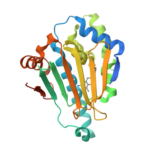
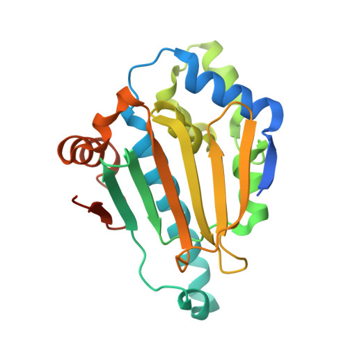
    4U93, 4W7T

  • PubMed Abstract: 

    Utilizing structure-based drug design, a novel dihydropyridopyrimidinone series which exhibited potent Hsp90 inhibition, good pharmacokinetics upon oral administration, and an excellent pharmacokinetic/pharmacodynamic relationship in vivo was developed from a commercial hit. The exploration of this series led to the selection of NVP-HSP990 as a development candidate.


  • Organizational Affiliation

    Global Discovery Chemistry/Oncology & Exploratory Chemistry, Novartis Institutes for Biomedical Research , 5300 Chiron Way, Emeryville, California 94608, United States.


Macromolecules
Find similar proteins by:  (by identity cutoff)  |  3D Structure
Entity ID: 1
MoleculeChains Sequence LengthOrganismDetailsImage
Heat shock protein HSP 90-alpha236Homo sapiensMutation(s): 0 
EC: 3.6.4.10
UniProt & NIH Common Fund Data Resources
Find proteins for P07900 (Homo sapiens)
Explore P07900 
Go to UniProtKB:  P07900
PHAROS:  P07900
GTEx:  ENSG00000080824 
Entity Groups  
Sequence Clusters30% Identity50% Identity70% Identity90% Identity95% Identity100% Identity
UniProt GroupP07900
Sequence Annotations
Expand
  • Reference Sequence
Small Molecules
Ligands 1 Unique
IDChains Name / Formula / InChI Key2D Diagram3D Interactions
3JC
Query on 3JC

Download Ideal Coordinates CCD File 
B [auth A](7S)-2-amino-4-methyl-7-phenyl-7,8-dihydroquinazolin-5(6H)-one
C15 H15 N3 O
PHMJMYBSWXYOLM-NSHDSACASA-N
Experimental Data & Validation

Experimental Data

  • Method: X-RAY DIFFRACTION
  • Resolution: 1.80 Å
  • R-Value Free:  0.210 (Depositor), 0.200 (DCC) 
  • R-Value Work:  0.171 (Depositor), 0.170 (DCC) 
  • R-Value Observed: 0.173 (Depositor) 
Space Group: I 2 2 2
Unit Cell:
Length ( Å )Angle ( ˚ )
a = 64.095α = 90
b = 89.983β = 90
c = 97.985γ = 90
Software Package:
Software NamePurpose
PHENIXrefinement
EPMRphasing
PDB_EXTRACTdata extraction

Structure Validation

View Full Validation Report


Ligand Structure Quality Assessment 

Created with Raphaël 2.3.0Worse 01 BetterLigand structure goodness of fit to experimental dataBest fitted 3JCClick on this verticalbar to view details

Entry History 

Deposition Data

Revision History  (Full details and data files)

  • Version 1.0: 2014-11-26
    Type: Initial release
  • Version 1.1: 2023-12-27
    Changes: Data collection, Database references, Derived calculations, Other, Refinement description, Source and taxonomy
  • Version 1.2: 2024-04-03
    Changes: Refinement description