4PSR

Crystal Structure of alpha-L-fucosidase from Fusarium graminearum in the open form in complex with L-fucose


Experimental Data Snapshot

  • Method: X-RAY DIFFRACTION
  • Resolution: 1.38 Å
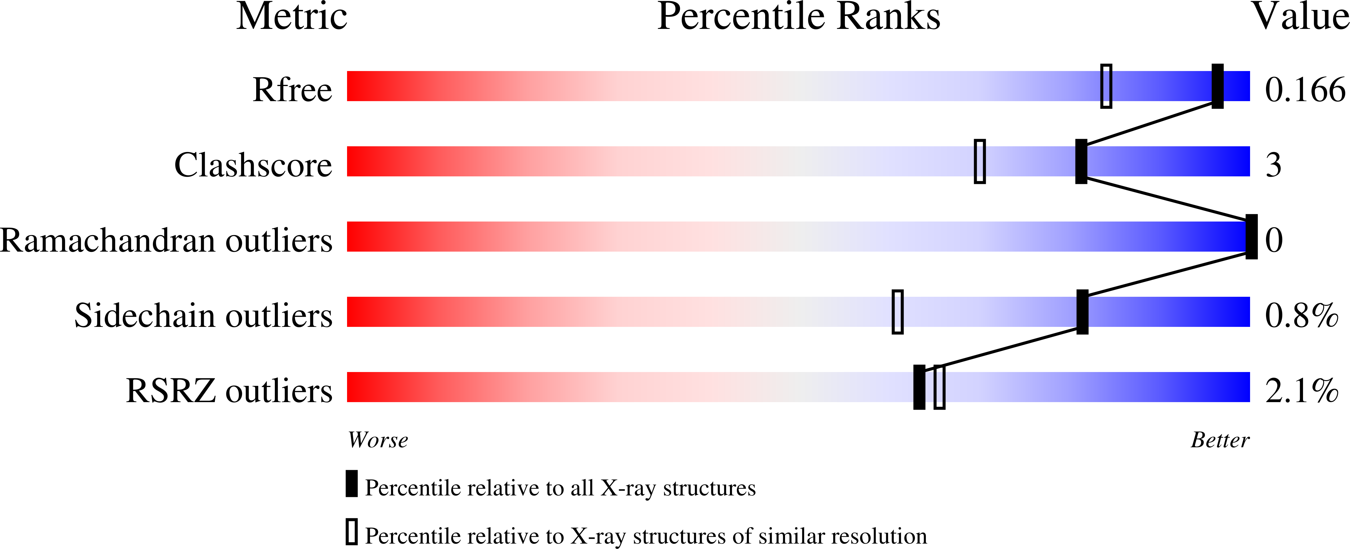
  • R-Value Free: 
    0.166 (Depositor), 0.170 (DCC) 
  • R-Value Work: 
    0.138 (Depositor), 0.140 (DCC) 
  • R-Value Observed: 
    0.140 (Depositor) 

Starting Model: experimental
View more details

wwPDB Validation   3D Report Full Report


Ligand Structure Quality Assessment 

Created with Raphaël 2.3.0Worse 01 BetterLigand structure goodness of fit to experimental dataBest fitted NAGClick on this verticalbar to view detailsBest fitted FULClick on this verticalbar to view details

This is version 2.2 of the entry. See complete history


Literature

Structure and Substrate Specificity of a Eukaryotic Fucosidase from Fusarium graminearum.

Cao, H.Walton, J.D.Brumm, P.Phillips, G.N.

(2014) J Biol Chem 289: 25624-25638

  • DOI: https://doi.org/10.1074/jbc.M114.583286
  • Primary Citation of Related Structures:  
    4NI3, 4PSP, 4PSR

  • PubMed Abstract: 

    The secreted glycoside hydrolase family 29 (GH29) α-L-fucosidase from plant pathogenic fungus Fusarium graminearum (FgFCO1) actively releases fucose from the xyloglucan fragment. We solved crystal structures of two active-site conformations, i.e. open and closed, of apoFgFCO1 and an open complex with product fucose at atomic resolution. The closed conformation supports catalysis by orienting the conserved general acid/base Glu-288 nearest the predicted glycosidic position, whereas the open conformation possibly represents an unreactive state with Glu-288 positioned away from the catalytic center. A flexible loop near the substrate binding site containing a non-conserved GGSFT sequence is ordered in the closed but not the open form. We also identified a novel C-terminal βγ-crystallin domain in FgFCO1 devoid of calcium binding motif whose homologous sequences are present in various glycoside hydrolase families. N-Glycosylated FgFCO1 adopts a monomeric state as verified by solution small angle x-ray scattering in contrast to reported multimeric fucosidases. Steady-state kinetics shows that FgFCO1 prefers α1,2 over α1,3/4 linkages and displays minimal activity with p-nitrophenyl fucoside with an acidic pH optimum of 4.6. Despite a retaining GH29 family fold, the overall specificity of FgFCO1 most closely resembles inverting GH95 α-fucosidase, which displays the highest specificity with two natural substrates harboring the Fucα1-2Gal glycosidic linkage, a xyloglucan-derived nonasaccharide, and 2'-fucosyllactose. Furthermore, FgFCO1 hydrolyzes H-disaccharide (lacking a +2 subsite sugar) at a rate 10(3)-fold slower than 2'-fucosyllactose. We demonstrated the structurally dynamic active site of FgFCO1 with flexible general acid/base Glu, a common feature shared by several bacterial GH29 fucosidases to various extents.


  • Organizational Affiliation

    From Rice University, Houston Texas 77005, Great Lakes Bioenergy Research Center, University of Wisconsin, Madison, Wisconsin 53706.


Macromolecules
Find similar proteins by:  (by identity cutoff)  |  3D Structure
Entity ID: 1
MoleculeChains Sequence LengthOrganismDetailsImage
Alpha-fucosidase GH29
A, B
585Fusarium graminearumMutation(s): 0 
Gene Names: FCO1
EC: 3.2.1.51
UniProt
Find proteins for J9UN47 (Gibberella zeae)
Explore J9UN47 
Go to UniProtKB:  J9UN47
Entity Groups  
Sequence Clusters30% Identity50% Identity70% Identity90% Identity95% Identity100% Identity
UniProt GroupJ9UN47
Glycosylation
Glycosylation Sites: 3
Sequence Annotations
Expand
  • Reference Sequence
Oligosaccharides

Help

Entity ID: 2
MoleculeChains Length2D Diagram Glycosylation3D Interactions
alpha-D-mannopyranose-(1-2)-alpha-D-mannopyranose-(1-2)-alpha-D-mannopyranose-(1-3)-[alpha-D-mannopyranose-(1-6)]beta-D-mannopyranose-(1-4)-2-acetamido-2-deoxy-beta-D-glucopyranose-(1-4)-2-acetamido-2-deoxy-beta-D-glucopyranose
C
7N-Glycosylation
Glycosylation Resources
GlyTouCan:  G46836GH
GlyCosmos:  G46836GH
GlyGen:  G46836GH
Entity ID: 3
MoleculeChains Length2D Diagram Glycosylation3D Interactions
alpha-D-mannopyranose-(1-2)-alpha-D-mannopyranose-(1-2)-alpha-D-mannopyranose-(1-3)-beta-D-mannopyranose-(1-4)-2-acetamido-2-deoxy-beta-D-glucopyranose-(1-4)-2-acetamido-2-deoxy-beta-D-glucopyranose
D
6N-Glycosylation
Glycosylation Resources
GlyTouCan:  G01760ZU
GlyCosmos:  G01760ZU
GlyGen:  G01760ZU
Small Molecules
Ligands 5 Unique
IDChains Name / Formula / InChI Key2D Diagram3D Interactions
NAG
Query on NAG

Download Ideal Coordinates CCD File 
E [auth A],
F [auth A],
N [auth B]
2-acetamido-2-deoxy-beta-D-glucopyranose
C8 H15 N O6
OVRNDRQMDRJTHS-FMDGEEDCSA-N
FUL
Query on FUL

Download Ideal Coordinates CCD File 
M [auth A],
V [auth B]
beta-L-fucopyranose
C6 H12 O5
SHZGCJCMOBCMKK-KGJVWPDLSA-N
TRS
Query on TRS

Download Ideal Coordinates CCD File 
G [auth A]
H [auth A]
I [auth A]
O [auth B]
P [auth B]
2-AMINO-2-HYDROXYMETHYL-PROPANE-1,3-DIOL
C4 H12 N O3
LENZDBCJOHFCAS-UHFFFAOYSA-O
GOL
Query on GOL

Download Ideal Coordinates CCD File 
K [auth A],
L [auth A],
T [auth B],
U [auth B]
GLYCEROL
C3 H8 O3
PEDCQBHIVMGVHV-UHFFFAOYSA-N
NA
Query on NA

Download Ideal Coordinates CCD File 
J [auth A],
R [auth B],
S [auth B]
SODIUM ION
Na
FKNQFGJONOIPTF-UHFFFAOYSA-N
Experimental Data & Validation

Experimental Data

  • Method: X-RAY DIFFRACTION
  • Resolution: 1.38 Å
  • R-Value Free:  0.166 (Depositor), 0.170 (DCC) 
  • R-Value Work:  0.138 (Depositor), 0.140 (DCC) 
  • R-Value Observed: 0.140 (Depositor) 
Space Group: P 1
Unit Cell:
Length ( Å )Angle ( ˚ )
a = 54.153α = 105.69
b = 75.989β = 107.32
c = 80.97γ = 106.93
Software Package:
Software NamePurpose
DENZOdata reduction
SCALEPACKdata scaling
PHENIXrefinement
PDB_EXTRACTdata extraction
MD2data collection
HKL-2000data reduction
HKL-2000data scaling
PHASERphasing

Structure Validation

View Full Validation Report


Ligand Structure Quality Assessment 

Created with Raphaël 2.3.0Worse 01 BetterLigand structure goodness of fit to experimental dataBest fitted NAGClick on this verticalbar to view detailsBest fitted FULClick on this verticalbar to view details

Entry History 

Deposition Data

Revision History  (Full details and data files)

  • Version 1.0: 2014-03-19
    Type: Initial release
  • Version 1.1: 2014-09-17
    Changes: Database references
  • Version 1.2: 2014-10-01
    Changes: Database references
  • Version 1.3: 2017-11-22
    Changes: Refinement description
  • Version 2.0: 2020-07-29
    Type: Remediation
    Reason: Carbohydrate remediation
    Changes: Advisory, Atomic model, Data collection, Derived calculations, Structure summary
  • Version 2.1: 2023-09-20
    Changes: Data collection, Database references, Refinement description, Structure summary
  • Version 2.2: 2024-10-30
    Changes: Structure summary