4N02 | pdb_00004n02

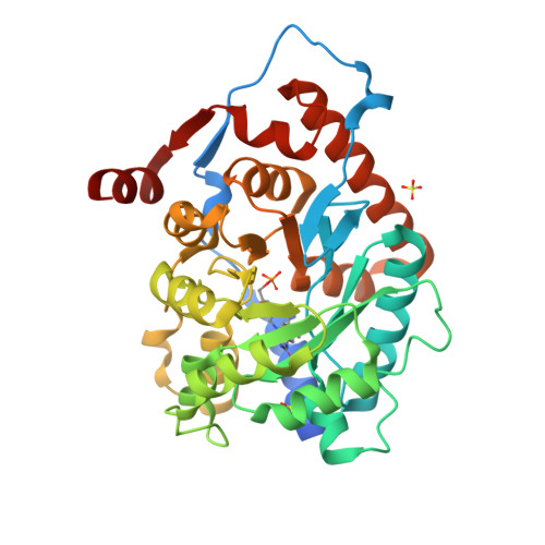
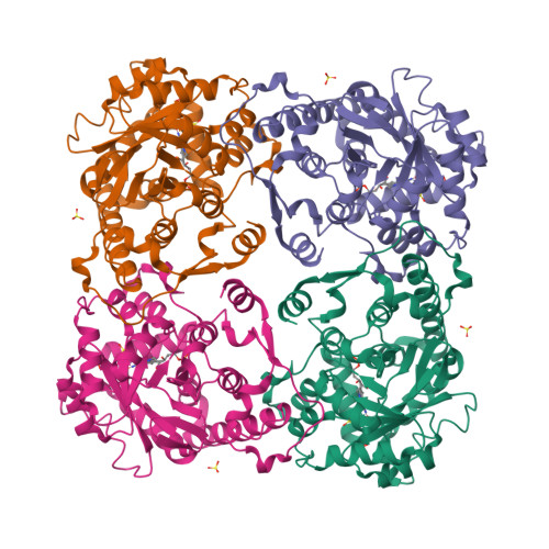
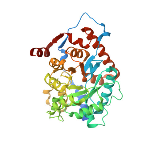
Type 2 IDI from S. pneumoniae


Experimental Data Snapshot

  • Method: X-RAY DIFFRACTION
  • Resolution: 1.40 Å
  • R-Value Free: 
    0.159 (Depositor), 0.150 (DCC) 
  • R-Value Work: 
    0.139 (Depositor), 0.130 (DCC) 
  • R-Value Observed: 
    0.140 (Depositor) 

wwPDB Validation   3D Report Full Report


Ligand Structure Quality Assessment 

Created with Raphaël 2.3.0Worse 01 BetterLigand structure goodness of fit to experimental dataBest fitted FNRClick on this verticalbar to view details

This is version 1.2 of the entry. See complete history


Literature

Determination of Kinetics and the Crystal Structure of a Novel Type 2 Isopentenyl Diphosphate: Dimethylallyl Diphosphate Isomerase from Streptococcus pneumoniae.

de Ruyck, J.Janczak, M.W.Neti, S.S.Rothman, S.C.Schubert, H.L.Cornish, R.M.Matagne, A.Wouters, J.Poulter, C.D.

(2014) Chembiochem 15: 1452-1458

  • DOI: https://doi.org/10.1002/cbic.201402046
  • Primary Citation of Related Structures:  
    4N02

  • PubMed Abstract: 

    Isopentenyl diphosphate isomerase (IDI) is a key enzyme in the isoprenoid biosynthetic pathway and is required for all organisms that synthesize isoprenoid metabolites from mevalonate. Type 1 IDI (IDI-1) is a metalloprotein that is found in eukaryotes, whereas the type 2 isoform (IDI-2) is a flavoenzyme found in bacteria that is completely absent from human. IDI-2 from the pathogenic bacterium Streptococcus pneumoniae was recombinantly expressed in Escherichia coli. Steady-state kinetic studies of the enzyme indicated that FMNH2 (KM =0.3 μM) bound before isopentenyl diphosphate (KM =40 μM) in an ordered binding mechanism. An X-ray crystal structure at 1.4 Å resolution was obtained for the holoenzyme in the closed conformation with a reduced flavin cofactor and two sulfate ions in the active site. These results helped to further approach the enzymatic mechanism of IDI-2 and, thus, open new possibilities for the rational design of antibacterial compounds against sequence-similar and structure-related pathogens such as Enterococcus faecalis or Staphylococcus aureus.


  • Organizational Affiliation

    Department of Chemistry, University of Utah, 315 South 1400 East RM 2020, Salt Lake City, Utah 84112 (USA); Department of Chemistry, UNamur, 61 rue de Bruxelles, 5000 Namur (Belgium).


Macromolecules
Find similar proteins by:  (by identity cutoff)  |  3D Structure
Entity ID: 1
MoleculeChains Sequence LengthOrganismDetailsImage
Isopentenyl-diphosphate delta-isomerase357Streptococcus pneumoniae CGSP14Mutation(s): 0 
EC: 5.3.3.2
UniProt
Find proteins for B2ILS5 (Streptococcus pneumoniae (strain CGSP14))
Explore B2ILS5 
Go to UniProtKB:  B2ILS5
Entity Groups  
Sequence Clusters30% Identity50% Identity70% Identity90% Identity95% Identity100% Identity
UniProt GroupB2ILS5
Sequence Annotations
Expand
  • Reference Sequence
Experimental Data & Validation

Experimental Data

  • Method: X-RAY DIFFRACTION
  • Resolution: 1.40 Å
  • R-Value Free:  0.159 (Depositor), 0.150 (DCC) 
  • R-Value Work:  0.139 (Depositor), 0.130 (DCC) 
  • R-Value Observed: 0.140 (Depositor) 
Space Group: I 4
Unit Cell:
Length ( Å )Angle ( ˚ )
a = 131.329α = 90
b = 131.329β = 90
c = 59.412γ = 90
Software Package:
Software NamePurpose
PHENIXrefinement

Structure Validation

View Full Validation Report


Ligand Structure Quality Assessment 

Created with Raphaël 2.3.0Worse 01 BetterLigand structure goodness of fit to experimental dataBest fitted FNRClick on this verticalbar to view details

Entry History 

Deposition Data

Revision History  (Full details and data files)

  • Version 1.0: 2014-07-02
    Type: Initial release
  • Version 1.1: 2014-07-16
    Changes: Database references
  • Version 1.2: 2024-02-28
    Changes: Data collection, Database references, Derived calculations