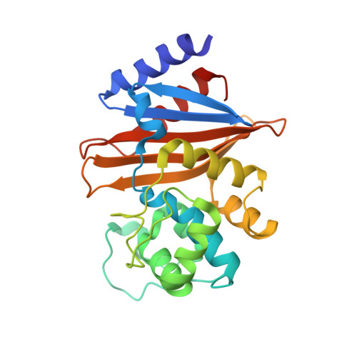
4JF4

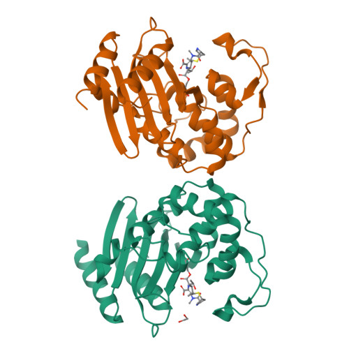
OXA-23 meropenem complex


Experimental Data Snapshot

  • Method: X-RAY DIFFRACTION
  • Resolution: 2.14 Å
  • R-Value Free: 0.254 
  • R-Value Work: 0.199 
  • R-Value Observed: 0.202 

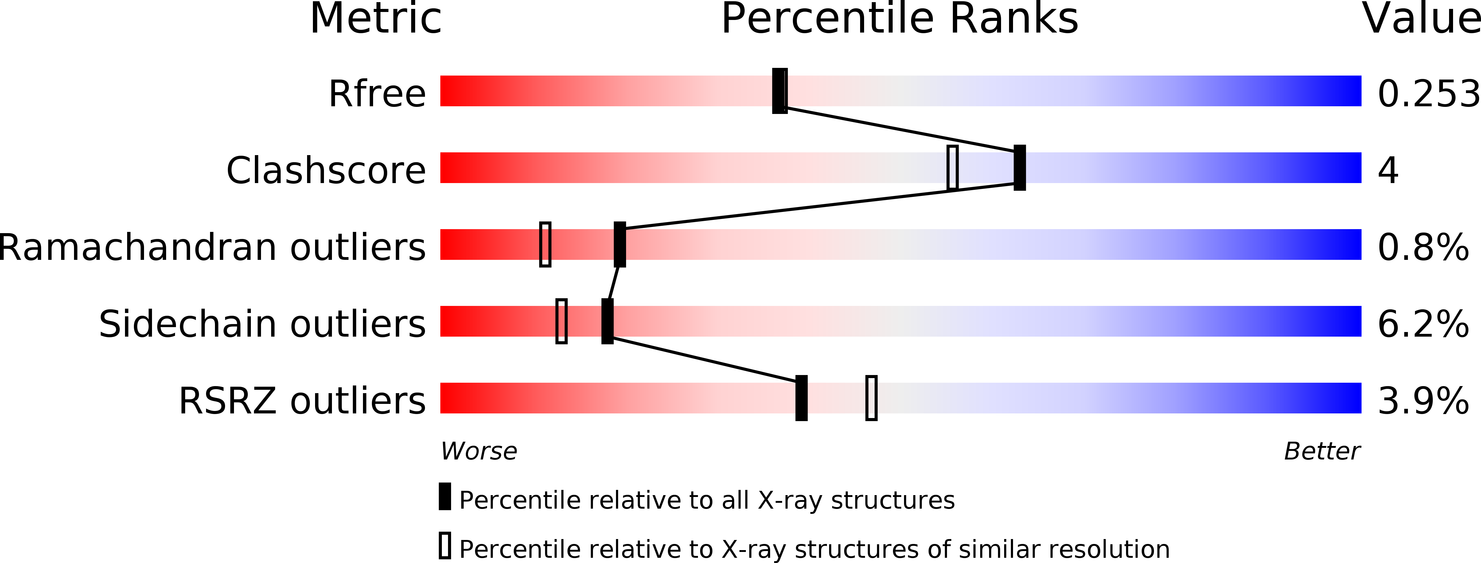
wwPDB Validation   3D Report Full Report


Ligand Structure Quality Assessment 


This is version 1.2 of the entry. See complete history


Literature

Structural Basis for Carbapenemase Activity of the OXA-23 beta-Lactamase from Acinetobacter baumannii.

Smith, C.A.Antunes, N.T.Stewart, N.K.Toth, M.Kumarasiri, M.Chang, M.Mobashery, S.Vakulenko, S.B.

(2013) Chem Biol 20: 1107-1115

  • DOI: https://doi.org/10.1016/j.chembiol.2013.07.015
  • Primary Citation of Related Structures:  
    4JF4, 4JF5, 4JF6

  • PubMed Abstract: 

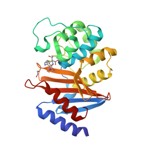
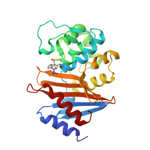
    Dissemination of Acinetobacter baumannii strains harboring class D β-lactamases producing resistance to carbapenem antibiotics severely limits our ability to treat deadly Acinetobacter infections. Susceptibility determination in the A. baumannii background and kinetic studies with a homogeneous preparation of OXA-23 β-lactamase, the major carbapenemase present in A. baumannii, document the ability of this enzyme to manifest resistance to last-resort carbapenem antibiotics. We also report three X-ray structures of OXA-23: apo OXA-23 at two different pH values, and wild-type OXA-23 in complex with meropenem, a carbapenem substrate. The structures and dynamics simulations reveal an important role for Leu166, whose motion regulates the access of a hydrolytic water molecule to the acyl-enzyme species in imparting carbapenemase activity.


  • Organizational Affiliation

    Stanford Synchrotron Radiation Lightsource, Stanford University, Menlo Park, CA 94025, USA. Electronic address: [email protected].


Macromolecules
Find similar proteins by:  (by identity cutoff)  |  3D Structure
Entity ID: 1
MoleculeChains Sequence LengthOrganismDetailsImage
Beta-lactamase
A, B
243Acinetobacter baumanniiMutation(s): 0 
Gene Names: ari-1bla(OXA-23)bla-OXA-23blaOXA-23OXA-23oxa23
EC: 3.5.2.6
UniProt
Find proteins for Q9L4P2 (Acinetobacter baumannii)
Explore Q9L4P2 
Go to UniProtKB:  Q9L4P2
Entity Groups  
Sequence Clusters30% Identity50% Identity70% Identity90% Identity95% Identity100% Identity
UniProt GroupQ9L4P2
Sequence Annotations
Expand
  • Reference Sequence
Small Molecules
Ligands 2 Unique
IDChains Name / Formula / InChI Key2D Diagram3D Interactions
MER
Query on MER

Download Ideal Coordinates CCD File 
D [auth A],
E [auth B]
(4R,5S)-3-{[(3S,5S)-5-(dimethylcarbamoyl)pyrrolidin-3-yl]sulfanyl}-5-[(2S,3R)-3-hydroxy-1-oxobutan-2-yl]-4-methyl-4,5-d ihydro-1H-pyrrole-2-carboxylic acid
C17 H27 N3 O5 S
DYQHXZPAIVAJRU-HTXLXMOSSA-N
EDO
Query on EDO

Download Ideal Coordinates CCD File 
C [auth A]1,2-ETHANEDIOL
C2 H6 O2
LYCAIKOWRPUZTN-UHFFFAOYSA-N
Experimental Data & Validation

Experimental Data

  • Method: X-RAY DIFFRACTION
  • Resolution: 2.14 Å
  • R-Value Free: 0.254 
  • R-Value Work: 0.199 
  • R-Value Observed: 0.202 
  • Space Group: P 1 21 1
Unit Cell:
Length ( Å )Angle ( ˚ )
a = 49.467α = 90
b = 44.533β = 94.93
c = 131.367γ = 90
Software Package:
Software NamePurpose
Blu-Icedata collection
MOLREPphasing
PHENIXrefinement
XDSdata reduction
SCALAdata scaling

Structure Validation

View Full Validation Report


Ligand Structure Quality Assessment 


Entry History 

Deposition Data

Revision History  (Full details and data files)

  • Version 1.0: 2013-09-25
    Type: Initial release
  • Version 1.1: 2013-10-09
    Changes: Database references
  • Version 1.2: 2024-10-09
    Changes: Data collection, Database references, Derived calculations, Structure summary