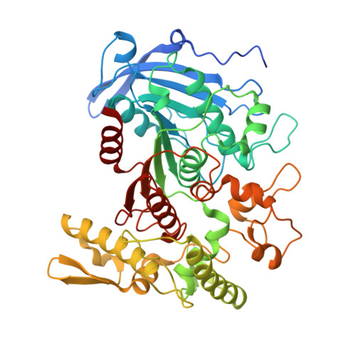
4J0I

Tannin acyl hydrolase in complex with 3,4-dihydroxybenzoate


Experimental Data Snapshot

  • Method: X-RAY DIFFRACTION
  • Resolution: 1.75 Å
  • R-Value Free: 0.188 
  • R-Value Work: 0.155 
  • R-Value Observed: 0.156 

wwPDB Validation   3D Report Full Report


Ligand Structure Quality Assessment 


This is version 1.2 of the entry. See complete history


Literature

Crystal structure of tannase from Lactobacillus plantarum.

Ren, B.Wu, M.Wang, Q.Peng, X.Wen, H.McKinstry, W.J.Chen, Q.

(2013) J Mol Biol 425: 2737-2751

  • DOI: https://doi.org/10.1016/j.jmb.2013.04.032
  • Primary Citation of Related Structures:  
    4J0C, 4J0D, 4J0G, 4J0H, 4J0I, 4J0J, 4J0K, 4JUI

  • PubMed Abstract: 

    Tannins are water-soluble polyphenolic compounds in plants. Hydrolyzable tannins are derivatives of gallic acid (3,4,5-trihydroxybenzoic acid) or its meta-depsidic forms that are esterified to polyol, catechin, or triterpenoid units. Tannases are a family of esterases that catalyze the hydrolysis of the galloyl ester bond in hydrolyzable tannins to release gallic acid. The enzymes have found wide applications in food, feed, beverage, pharmaceutical, and chemical industries since their discovery more than a century ago, although little is known about them at the molecular level, including the details of the catalytic and substrate binding sites. Here, we report the first three-dimensional structure of a tannase from Lactobacillus plantarum. The enzyme displays an α/β structure, featured by a large cap domain inserted into the classical serine hydrolase fold. A catalytic triad was identified in the structure, which is composed of Ser163, His451, and Asp419. During the binding of gallic acid, the carboxyl group of the molecule forges hydrogen-bonding interactions with the catalytic triad of the enzyme while the three hydroxyl groups make contacts with Asp421, Lys343, and Glu357 to form another hydrogen-bonding network. Mutagenesis studies demonstrated that these residues are indispensable for the activity of the enzyme. Structural studies of the enzyme in complex with a number of substrates indicated that the interactions at the galloyl binding site are the determinant force for the binding of substrates. The single galloyl binding site is responsible for the esterase and depsidase activities of the enzyme.


  • Organizational Affiliation

    Materials Science and Engineering, CSIRO, 343 Royal Parade, Parkville, VIC 3052, Australia. [email protected]


Macromolecules
Find similar proteins by:  (by identity cutoff)  |  3D Structure
Entity ID: 1
MoleculeChains Sequence LengthOrganismDetailsImage
Tannase
A, B
491Lactiplantibacillus plantarumMutation(s): 0 
Gene Names: tanLpl
UniProt
Find proteins for B3Y018 (Lactiplantibacillus plantarum)
Explore B3Y018 
Go to UniProtKB:  B3Y018
Entity Groups  
Sequence Clusters30% Identity50% Identity70% Identity90% Identity95% Identity100% Identity
UniProt GroupB3Y018
Sequence Annotations
Expand
  • Reference Sequence
Small Molecules
Ligands 3 Unique
IDChains Name / Formula / InChI Key2D Diagram3D Interactions
PG4
Query on PG4

Download Ideal Coordinates CCD File 
G [auth B]TETRAETHYLENE GLYCOL
C8 H18 O5
UWHCKJMYHZGTIT-UHFFFAOYSA-N
DHB
Query on DHB

Download Ideal Coordinates CCD File 
C [auth A],
F [auth B]
3,4-DIHYDROXYBENZOIC ACID
C7 H6 O4
YQUVCSBJEUQKSH-UHFFFAOYSA-N
PEG
Query on PEG

Download Ideal Coordinates CCD File 
D [auth A]
E [auth A]
H [auth B]
I [auth B]
J [auth B]
D [auth A],
E [auth A],
H [auth B],
I [auth B],
J [auth B],
K [auth B],
L [auth B]
DI(HYDROXYETHYL)ETHER
C4 H10 O3
MTHSVFCYNBDYFN-UHFFFAOYSA-N
Experimental Data & Validation

Experimental Data

  • Method: X-RAY DIFFRACTION
  • Resolution: 1.75 Å
  • R-Value Free: 0.188 
  • R-Value Work: 0.155 
  • R-Value Observed: 0.156 
  • Space Group: P 1
Unit Cell:
Length ( Å )Angle ( ˚ )
a = 46.578α = 70.35
b = 62.852β = 86.05
c = 83.853γ = 79.48
Software Package:
Software NamePurpose
Blu-Icedata collection
PHENIXmodel building
PHENIXrefinement
XDSdata reduction
SCALAdata scaling
PHENIXphasing

Structure Validation

View Full Validation Report


Ligand Structure Quality Assessment 


Entry History 

Deposition Data

Revision History  (Full details and data files)

  • Version 1.0: 2013-05-22
    Type: Initial release
  • Version 1.1: 2022-08-24
    Changes: Database references, Derived calculations
  • Version 1.2: 2024-05-29
    Changes: Data collection