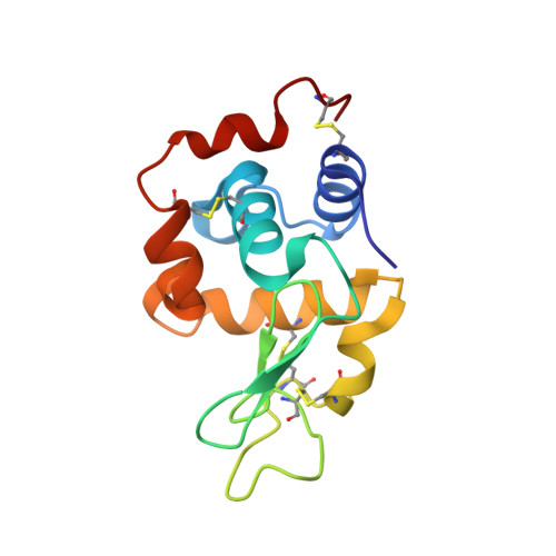
4HTQ

Mitigation of X-ray damage in macromolecular crystallography by submicrometer line focusing; total dose 6.70 x 10e+11 X-ray photons


Experimental Data Snapshot

  • Method: X-RAY DIFFRACTION
  • Resolution: 1.40 Å
  • R-Value Free: 0.163 
  • R-Value Work: 0.130 
  • R-Value Observed: 0.132 

wwPDB Validation   3D Report Full Report


This is version 1.3 of the entry. See complete history


Literature

Mitigation of X-ray damage in macromolecular crystallography by submicrometre line focusing.

Finfrock, Y.Z.Stern, E.A.Alkire, R.W.Kas, J.J.Evans-Lutterodt, K.Stein, A.Duke, N.Lazarski, K.Joachimiak, A.

(2013) Acta Crystallogr D Biol Crystallogr 69: 1463-1469

  • DOI: https://doi.org/10.1107/S0907444913009335
  • Primary Citation of Related Structures:  
    4HTK, 4HTN, 4HTQ

  • PubMed Abstract: 

    Reported here are measurements of the penetration depth and spatial distribution of photoelectron (PE) damage excited by 18.6 keV X-ray photons in a lysozyme crystal with a vertical submicrometre line-focus beam of 0.7 µm full-width half-maximum (FWHM). The experimental results determined that the penetration depth of PEs is 5 ± 0.5 µm with a monotonically decreasing spatial distribution shape, resulting in mitigation of diffraction signal damage. This does not agree with previous theoretical predication that the mitigation of damage requires a peak of damage outside the focus. A new improved calculation provides some qualitative agreement with the experimental results, but significant errors still remain. The mitigation of radiation damage by line focusing was measured experimentally by comparing the damage in the X-ray-irradiated regions of the submicrometre focus with the large-beam case under conditions of equal exposure and equal volumes of the protein crystal, and a mitigation factor of 4.4 ± 0.4 was determined. The mitigation of radiation damage is caused by spatial separation of the dominant PE radiation-damage component from the crystal region of the line-focus beam that contributes the diffraction signal. The diffraction signal is generated by coherent scattering of incident X-rays (which introduces no damage), while the overwhelming proportion of damage is caused by PE emission as X-ray photons are absorbed.


  • Organizational Affiliation

    Physics Department, University of Washington, Seattle, WA 98195, USA.


Macromolecules
Find similar proteins by:  (by identity cutoff)  |  3D Structure
Entity ID: 1
MoleculeChains Sequence LengthOrganismDetailsImage
Lysozyme C129Gallus gallusMutation(s): 0 
EC: 3.2.1.17
UniProt
Find proteins for P00698 (Gallus gallus)
Explore P00698 
Go to UniProtKB:  P00698
Entity Groups  
Sequence Clusters30% Identity50% Identity70% Identity90% Identity95% Identity100% Identity
UniProt GroupP00698
Sequence Annotations
Expand
  • Reference Sequence
Small Molecules
Ligands 3 Unique
IDChains Name / Formula / InChI Key2D Diagram3D Interactions
EDO
Query on EDO

Download Ideal Coordinates CCD File 
K [auth A],
L [auth A],
M [auth A]
1,2-ETHANEDIOL
C2 H6 O2
LYCAIKOWRPUZTN-UHFFFAOYSA-N
CL
Query on CL

Download Ideal Coordinates CCD File 
B [auth A]
C [auth A]
D [auth A]
E [auth A]
F [auth A]
B [auth A],
C [auth A],
D [auth A],
E [auth A],
F [auth A],
G [auth A],
H [auth A],
I [auth A]
CHLORIDE ION
Cl
VEXZGXHMUGYJMC-UHFFFAOYSA-M
NA
Query on NA

Download Ideal Coordinates CCD File 
J [auth A]SODIUM ION
Na
FKNQFGJONOIPTF-UHFFFAOYSA-N
Experimental Data & Validation

Experimental Data

  • Method: X-RAY DIFFRACTION
  • Resolution: 1.40 Å
  • R-Value Free: 0.163 
  • R-Value Work: 0.130 
  • R-Value Observed: 0.132 
  • Space Group: P 43 21 2
Unit Cell:
Length ( Å )Angle ( ˚ )
a = 78.922α = 90
b = 78.922β = 90
c = 36.868γ = 90
Software Package:
Software NamePurpose
DENZOdata reduction
SCALEPACKdata scaling
MOLREPphasing
PHENIXrefinement
PDB_EXTRACTdata extraction
SBC-Collectdata collection

Structure Validation

View Full Validation Report


Entry History 

Deposition Data

Revision History  (Full details and data files)

  • Version 1.0: 2013-05-15
    Type: Initial release
  • Version 1.1: 2013-08-28
    Changes: Database references
  • Version 1.2: 2017-11-15
    Changes: Refinement description
  • Version 1.3: 2024-11-27
    Changes: Data collection, Database references, Derived calculations, Structure summary