4HEF | pdb_00004hef

Structure of avibactam bound to Pseudomonas aeruginosa AmpC


Experimental Data Snapshot

  • Method: X-RAY DIFFRACTION
  • Resolution: 1.86 Å
  • R-Value Free: 
    0.202 (Depositor), 0.220 (DCC) 
  • R-Value Work: 
    0.174 (Depositor), 0.190 (DCC) 
  • R-Value Observed: 
    0.176 (Depositor) 

wwPDB Validation   3D Report Full Report


Ligand Structure Quality Assessment 

Created with Raphaël 2.3.0Worse 01 BetterLigand structure goodness of fit to experimental dataBest fitted NXLClick on this verticalbar to view details

This is version 1.4 of the entry. See complete history


Literature

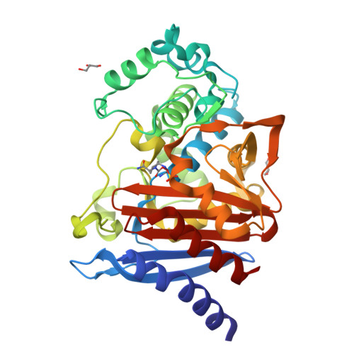
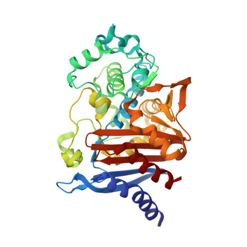
Structural Insight into Potent Broad-Spectrum Inhibition with Reversible Recyclization Mechanism: Avibactam in Complex with CTX-M-15 and Pseudomonas aeruginosa AmpC beta-Lactamases

Lahiri, S.D.Mangani, S.Durand-Reville, T.Benvenuti, M.De Luca, F.Sanyal, G.Docquier, J.D.

(2013) Antimicrob Agents Chemother 57: 2496-2505

  • DOI: https://doi.org/10.1128/AAC.02247-12
  • Primary Citation of Related Structures:  
    4GZB, 4HBT, 4HBU, 4HEF

  • PubMed Abstract: 

    Although β-lactams have been the most effective class of antibacterial agents used in clinical practice for the past half century, their effectiveness on Gram-negative bacteria has been eroded due to the emergence and spread of β-lactamase enzymes that are not affected by currently marketed β-lactam/β-lactamase inhibitor combinations. Avibactam is a novel, covalent, non-β-lactam β-lactamase inhibitor presently in clinical development in combination with either ceftaroline or ceftazidime. In vitro studies show that avibactam may restore the broad-spectrum activity of cephalosporins against class A, class C, and some class D β-lactamases. Here we describe the structures of two clinically important β-lactamase enzymes bound to avibactam, the class A CTX-M-15 extended-spectrum β-lactamase and the class C Pseudomonas aeruginosa AmpC β-lactamase, which together provide insight into the binding modes for the respective enzyme classes. The structures reveal similar binding modes in both enzymes and thus provide a rationale for the broad-spectrum inhibitory activity of avibactam. Identification of the key residues surrounding the binding pocket allows for a better understanding of the potency of this scaffold. Finally, avibactam has recently been shown to be a reversible inhibitor, and the structures provide insights into the mechanism of avibactam recyclization. Analysis of the ultra-high-resolution CTX-M-15 structure suggests how the deacylation mechanism favors recyclization over hydrolysis.


  • Organizational Affiliation

    Infection Biosciences, AstraZeneca R&D Boston, Waltham, Massachusetts, USA.


Macromolecules
Find similar proteins by:  (by identity cutoff)  |  3D Structure
Entity ID: 1
MoleculeChains Sequence LengthOrganismDetailsImage
Beta-lactamase360Pseudomonas aeruginosa PAO1Mutation(s): 0 
Gene Names: ampCPA4110
EC: 3.5.2.6
UniProt
Find proteins for P24735 (Pseudomonas aeruginosa (strain ATCC 15692 / DSM 22644 / CIP 104116 / JCM 14847 / LMG 12228 / 1C / PRS 101 / PAO1))
Explore P24735 
Go to UniProtKB:  P24735
Entity Groups  
Sequence Clusters30% Identity50% Identity70% Identity90% Identity95% Identity100% Identity
UniProt GroupP24735
Sequence Annotations
Expand
  • Reference Sequence
Experimental Data & Validation

Experimental Data

  • Method: X-RAY DIFFRACTION
  • Resolution: 1.86 Å
  • R-Value Free:  0.202 (Depositor), 0.220 (DCC) 
  • R-Value Work:  0.174 (Depositor), 0.190 (DCC) 
  • R-Value Observed: 0.176 (Depositor) 
Space Group: P 21 21 21
Unit Cell:
Length ( Å )Angle ( ˚ )
a = 44.88α = 90
b = 71.2β = 90
c = 106.25γ = 90
Software Package:
Software NamePurpose
REFMACrefinement
PDB_EXTRACTdata extraction
CrystalCleardata collection
d*TREKdata reduction
SCALAdata scaling
AMoREphasing
BUSTERrefinement

Structure Validation

View Full Validation Report


Ligand Structure Quality Assessment 

Created with Raphaël 2.3.0Worse 01 BetterLigand structure goodness of fit to experimental dataBest fitted NXLClick on this verticalbar to view details

Entry History 

Deposition Data

  • Released Date: 2013-06-05 
  • Deposition Author(s): Lahiri, S.D.

Revision History  (Full details and data files)

  • Version 1.0: 2013-06-05
    Type: Initial release
  • Version 1.1: 2015-06-03
    Changes: Non-polymer description
  • Version 1.2: 2018-02-07
    Changes: Experimental preparation
  • Version 1.3: 2020-09-16
    Changes: Database references, Derived calculations, Structure summary
  • Version 1.4: 2024-10-30
    Changes: Data collection, Database references, Structure summary