4F4O

Structure of the Haptoglobin-Haemoglobin Complex


Experimental Data Snapshot

  • Method: X-RAY DIFFRACTION
  • Resolution: 2.90 Å
  • R-Value Free: 0.229 
  • R-Value Work: 0.211 
  • R-Value Observed: 0.212 

Starting Models: experimental
View more details

wwPDB Validation   3D Report Full Report


Ligand Structure Quality Assessment 


This is version 2.3 of the entry. See complete history


Literature

Structure of the haptoglobin-haemoglobin complex.

Andersen, C.B.Torvund-Jensen, M.Nielsen, M.J.de Oliveira, C.L.Hersleth, H.P.Andersen, N.H.Pedersen, J.S.Andersen, G.R.Moestrup, S.K.

(2012) Nature 489: 456-459

  • DOI: https://doi.org/10.1038/nature11369
  • Primary Citation of Related Structures:  
    4F4O

  • PubMed Abstract: 

    Red cell haemoglobin is the fundamental oxygen-transporting molecule in blood, but also a potentially tissue-damaging compound owing to its highly reactive haem groups. During intravascular haemolysis, such as in malaria and haemoglobinopathies, haemoglobin is released into the plasma, where it is captured by the protective acute-phase protein haptoglobin. This leads to formation of the haptoglobin-haemoglobin complex, which represents a virtually irreversible non-covalent protein-protein interaction. Here we present the crystal structure of the dimeric porcine haptoglobin-haemoglobin complex determined at 2.9 Å resolution. This structure reveals that haptoglobin molecules dimerize through an unexpected β-strand swap between two complement control protein (CCP) domains, defining a new fusion CCP domain structure. The haptoglobin serine protease domain forms extensive interactions with both the α- and β-subunits of haemoglobin, explaining the tight binding between haptoglobin and haemoglobin. The haemoglobin-interacting region in the αβ dimer is highly overlapping with the interface between the two αβ dimers that constitute the native haemoglobin tetramer. Several haemoglobin residues prone to oxidative modification after exposure to haem-induced reactive oxygen species are buried in the haptoglobin-haemoglobin interface, thus showing a direct protective role of haptoglobin. The haptoglobin loop previously shown to be essential for binding of haptoglobin-haemoglobin to the macrophage scavenger receptor CD163 (ref. 3) protrudes from the surface of the distal end of the complex, adjacent to the associated haemoglobin α-subunit. Small-angle X-ray scattering measurements of human haptoglobin-haemoglobin bound to the ligand-binding fragment of CD163 confirm receptor binding in this area, and show that the rigid dimeric complex can bind two receptors. Such receptor cross-linkage may facilitate scavenging and explain the increased functional affinity of multimeric haptoglobin-haemoglobin for CD163 (ref. 4).


  • Organizational Affiliation

    Department of Biomedicine, Aarhus University, 8000 Aarhus C, Denmark. [email protected]


Macromolecules
Find similar proteins by:  (by identity cutoff)  |  3D Structure
Entity ID: 1
MoleculeChains Sequence LengthOrganismDetailsImage
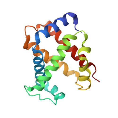
Hemoglobin subunit alpha
A, D, G, J
141Sus scrofaMutation(s): 0 
UniProt
Find proteins for P01965 (Sus scrofa)
Explore P01965 
Go to UniProtKB:  P01965
Entity Groups  
Sequence Clusters30% Identity50% Identity70% Identity90% Identity95% Identity100% Identity
UniProt GroupP01965
Sequence Annotations
Expand
  • Reference Sequence
Find similar proteins by:  (by identity cutoff)  |  3D Structure
Entity ID: 2
MoleculeChains Sequence LengthOrganismDetailsImage
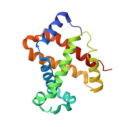
Hemoglobin subunit beta
B, E, H, K
146Sus scrofaMutation(s): 0 
UniProt
Find proteins for P02067 (Sus scrofa)
Explore P02067 
Go to UniProtKB:  P02067
Entity Groups  
Sequence Clusters30% Identity50% Identity70% Identity90% Identity95% Identity100% Identity
UniProt GroupP02067
Sequence Annotations
Expand
  • Reference Sequence
Find similar proteins by:  (by identity cutoff)  |  3D Structure
Entity ID: 3
MoleculeChains Sequence LengthOrganismDetailsImage
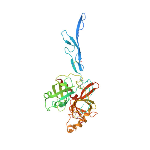
Haptoglobin
C, F, I, L
347Sus scrofaMutation(s): 0 
UniProt
Find proteins for Q8SPS7 (Sus scrofa)
Explore Q8SPS7 
Go to UniProtKB:  Q8SPS7
Entity Groups  
Sequence Clusters30% Identity50% Identity70% Identity90% Identity95% Identity100% Identity
UniProt GroupQ8SPS7
Glycosylation
Glycosylation Sites: 4
Sequence Annotations
Expand
  • Reference Sequence
Oligosaccharides

Help

Entity ID: 4
MoleculeChains Length2D Diagram Glycosylation3D Interactions
2-acetamido-2-deoxy-beta-D-glucopyranose-(1-4)-2-acetamido-2-deoxy-beta-D-glucopyranose
M, O, Q
2N-Glycosylation
Glycosylation Resources
GlyTouCan:  G42666HT
GlyCosmos:  G42666HT
GlyGen:  G42666HT
Entity ID: 5
MoleculeChains Length2D Diagram Glycosylation3D Interactions
alpha-L-fucopyranose-(1-6)-2-acetamido-2-deoxy-beta-D-glucopyranose
N, P, R, S
2N-Glycosylation
Glycosylation Resources
GlyTouCan:  G86851RC
GlyCosmos:  G86851RC
GlyGen:  G86851RC
Small Molecules
Ligands 3 Unique
IDChains Name / Formula / InChI Key2D Diagram3D Interactions
HEM
Query on HEM

Download Ideal Coordinates CCD File 
BA [auth E]
FA [auth G]
HA [auth H]
LA [auth J]
NA [auth K]
BA [auth E],
FA [auth G],
HA [auth H],
LA [auth J],
NA [auth K],
T [auth A],
V [auth B],
Z [auth D]
PROTOPORPHYRIN IX CONTAINING FE
C34 H32 Fe N4 O4
KABFMIBPWCXCRK-RGGAHWMASA-L
NAG
Query on NAG

Download Ideal Coordinates CCD File 
DA [auth F]
EA [auth F]
JA [auth I]
KA [auth I]
PA [auth L]
DA [auth F],
EA [auth F],
JA [auth I],
KA [auth I],
PA [auth L],
QA [auth L],
X [auth C],
Y [auth C]
2-acetamido-2-deoxy-beta-D-glucopyranose
C8 H15 N O6
OVRNDRQMDRJTHS-FMDGEEDCSA-N
OXY
Query on OXY

Download Ideal Coordinates CCD File 
AA [auth D]
CA [auth E]
GA [auth G]
IA [auth H]
MA [auth J]
AA [auth D],
CA [auth E],
GA [auth G],
IA [auth H],
MA [auth J],
OA [auth K],
U [auth A],
W [auth B]
OXYGEN MOLECULE
O2
MYMOFIZGZYHOMD-UHFFFAOYSA-N
Experimental Data & Validation

Experimental Data

  • Method: X-RAY DIFFRACTION
  • Resolution: 2.90 Å
  • R-Value Free: 0.229 
  • R-Value Work: 0.211 
  • R-Value Observed: 0.212 
  • Space Group: P 21 21 21
Unit Cell:
Length ( Å )Angle ( ˚ )
a = 72.88α = 90
b = 197.78β = 90
c = 322.07γ = 90
Software Package:
Software NamePurpose
PHASERphasing
PHENIXrefinement
XDSdata reduction
XDSdata scaling

Structure Validation

View Full Validation Report


Ligand Structure Quality Assessment 


Entry History 

Revision History  (Full details and data files)

  • Version 1.0: 2012-08-29
    Type: Initial release
  • Version 1.1: 2012-09-26
    Changes: Database references
  • Version 2.0: 2020-07-29
    Type: Remediation
    Reason: Carbohydrate remediation
    Changes: Atomic model, Data collection, Derived calculations, Source and taxonomy, Structure summary
  • Version 2.1: 2023-09-13
    Changes: Data collection, Database references, Refinement description, Structure summary
  • Version 2.2: 2024-03-13
    Changes: Source and taxonomy, Structure summary
  • Version 2.3: 2024-10-09
    Changes: Structure summary