4AYR

Structure of The GH47 processing alpha-1,2-mannosidase from Caulobacter strain K31 in complex with noeuromycin


Experimental Data Snapshot

  • Method: X-RAY DIFFRACTION
  • Resolution: 1.10 Å
  • R-Value Free: 0.102 
  • R-Value Work: 0.083 
  • R-Value Observed: 0.084 

Starting Model: experimental
View more details

wwPDB Validation   3D Report Full Report


Ligand Structure Quality Assessment 

Created with Raphaël 2.3.0Worse 01 BetterLigand structure goodness of fit to experimental dataBest fitted IFLClick on this verticalbar to view details

This is version 1.1 of the entry. See complete history


Literature

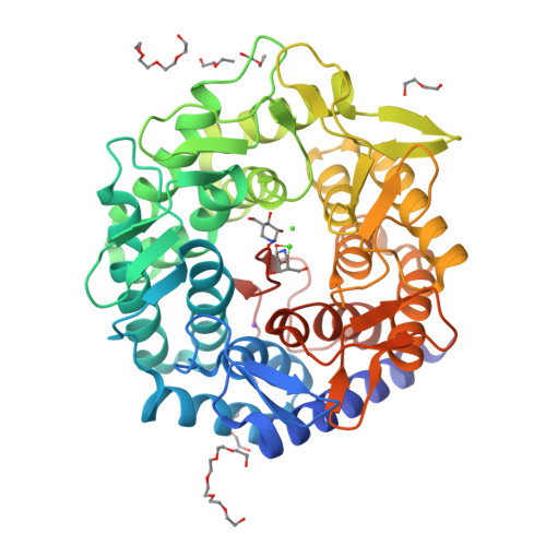
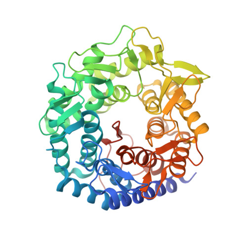
The Reaction Coordinate of a Bacterial Gh47 Alpha-Mannosidase: A Combined Quantum Mechanical and Structural Approach.

Thompson, A.J.Dabin, J.Iglesias-Fernandez, J.Ardevol, A.Dinev, Z.Williams, S.J.Bande, O.Siriwardena, A.Moreland, C.Hu, T.C.Smith, D.K.Gilbert, H.J.Rovira, C.Davies, G.J.

(2012) Angew Chem Int Ed Engl 51: 10997

  • DOI: https://doi.org/10.1002/anie.201205338
  • Primary Citation of Related Structures:  
    4AYO, 4AYP, 4AYQ, 4AYR

  • PubMed Abstract: 

    Mannosides in the southern hemisphere: Conformational analysis of enzymatic mannoside hydrolysis informs strategies for enzyme inhibition and inspires solutions to mannoside synthesis. Atomic resolution structures along the reaction coordinate of an inverting α-mannosidase show how the enzyme distorts the substrate and transition state. QM/MM calculations reveal how the free energy landscape of isolated α-D-mannose is molded on enzyme to only allow one conformationally accessible reaction coordinate.


  • Organizational Affiliation

    Department of Chemistry, University of York, Heslington, York, YO10 5DD, UK.


Macromolecules
Find similar proteins by:  (by identity cutoff)  |  3D Structure
Entity ID: 1
MoleculeChains Sequence LengthOrganismDetailsImage
MANNOSYL-OLIGOSACCHARIDE 1,2-ALPHA-MANNOSIDASE447Caulobacter sp. K31Mutation(s): 0 
EC: 3.2.1.113
UniProt
Find proteins for B0SWV2 (Caulobacter sp. (strain K31))
Explore B0SWV2 
Go to UniProtKB:  B0SWV2
Entity Groups  
Sequence Clusters30% Identity50% Identity70% Identity90% Identity95% Identity100% Identity
UniProt GroupB0SWV2
Sequence Annotations
Expand
  • Reference Sequence
Small Molecules
Ligands 4 Unique
IDChains Name / Formula / InChI Key2D Diagram3D Interactions
PE4
Query on PE4

Download Ideal Coordinates CCD File 
G [auth A],
H [auth A],
I [auth A],
J [auth A],
K [auth A]
2-{2-[2-(2-{2-[2-(2-ETHOXY-ETHOXY)-ETHOXY]-ETHOXY}-ETHOXY)-ETHOXY]-ETHOXY}-ETHANOL
C16 H34 O8
PJWQOENWHPEPKI-UHFFFAOYSA-N
IFL
Query on IFL

Download Ideal Coordinates CCD File 
E [auth A],
F [auth A]
(3S,4R,5R)-3,4-DIHYDROXY-5-(HYDROXYMETHYL)PIPERIDIN-2-ONE
C6 H11 N O4
ARBXEMIAJIJEQI-WDCZJNDASA-N
CA
Query on CA

Download Ideal Coordinates CCD File 
B [auth A],
C [auth A]
CALCIUM ION
Ca
BHPQYMZQTOCNFJ-UHFFFAOYSA-N
NA
Query on NA

Download Ideal Coordinates CCD File 
D [auth A]SODIUM ION
Na
FKNQFGJONOIPTF-UHFFFAOYSA-N
Binding Affinity Annotations 
IDSourceBinding Affinity
IFL PDBBind:  4AYR Kd: 99 (nM) from 1 assay(s)
Experimental Data & Validation

Experimental Data

  • Method: X-RAY DIFFRACTION
  • Resolution: 1.10 Å
  • R-Value Free: 0.102 
  • R-Value Work: 0.083 
  • R-Value Observed: 0.084 
  • Space Group: H 3
Unit Cell:
Length ( Å )Angle ( ˚ )
a = 143.919α = 90
b = 143.919β = 90
c = 50.186γ = 120
Software Package:
Software NamePurpose
REFMACrefinement
XDSdata reduction
SCALAdata scaling
MOLREPphasing

Structure Validation

View Full Validation Report


Ligand Structure Quality Assessment 

Created with Raphaël 2.3.0Worse 01 BetterLigand structure goodness of fit to experimental dataBest fitted IFLClick on this verticalbar to view details

Entry History 

Revision History  (Full details and data files)

  • Version 1.0: 2013-01-30
    Type: Initial release
  • Version 1.1: 2023-12-20
    Changes: Data collection, Database references, Derived calculations, Other, Refinement description