3W3N

Crystal structure of human TLR8 in complex with Resiquimod (R848) crystal form 3


Experimental Data Snapshot

  • Method: X-RAY DIFFRACTION
  • Resolution: 2.10 Å
  • R-Value Free: 
    0.233 (Depositor), 0.230 (DCC) 
  • R-Value Work: 
    0.200 (Depositor), 0.200 (DCC) 
  • R-Value Observed: 
    0.201 (Depositor) 

wwPDB Validation   3D Report Full Report


Ligand Structure Quality Assessment 

Created with Raphaël 2.3.0Worse 01 BetterLigand structure goodness of fit to experimental dataBest fitted RX8Click on this verticalbar to view detailsBest fitted NAGClick on this verticalbar to view details

This is version 2.2 of the entry. See complete history


Literature

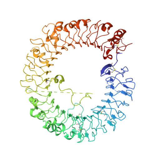
Structural reorganization of the Toll-like receptor 8 dimer induced by agonistic ligands

Tanji, H.Ohto, U.Shibata, T.Miyake, K.Shimizu, T.

(2013) Science 339: 1426-1429

  • DOI: https://doi.org/10.1126/science.1229159
  • Primary Citation of Related Structures:  
    3W3G, 3W3J, 3W3K, 3W3L, 3W3M, 3W3N

  • PubMed Abstract: 

    Toll-like receptor 7 (TLR7) and TLR8 recognize single-stranded RNA and initiate innate immune responses. Several synthetic agonists of TLR7-TLR8 display novel therapeutic potential; however, the molecular basis for ligand recognition and activation of signaling by TLR7 or TLR8 is largely unknown. In this study, the crystal structures of unliganded and ligand-induced activated human TLR8 dimers were elucidated. Ligand recognition was mediated by a dimerization interface formed by two protomers. Upon ligand stimulation, the TLR8 dimer was reorganized such that the two C termini were brought into proximity. The loop between leucine-rich repeat 14 (LRR14) and LRR15 was cleaved; however, the N- and C-terminal halves remained associated and contributed to ligand recognition and dimerization. Thus, ligand binding induces reorganization of the TLR8 dimer, which enables downstream signaling processes.


  • Organizational Affiliation

    Graduate School of Pharmaceutical Sciences, The University of Tokyo, Hongo, Bunkyo-ku, Tokyo 113-0033, Japan.


Macromolecules
Find similar proteins by:  (by identity cutoff)  |  3D Structure
Entity ID: 1
MoleculeChains Sequence LengthOrganismDetailsImage
Toll-like receptor 8
A, B
811Homo sapiensMutation(s): 0 
Gene Names: TLR8UNQ249/PRO286
UniProt & NIH Common Fund Data Resources
Find proteins for Q9NR97 (Homo sapiens)
Explore Q9NR97 
Go to UniProtKB:  Q9NR97
PHAROS:  Q9NR97
GTEx:  ENSG00000101916 
Entity Groups  
Sequence Clusters30% Identity50% Identity70% Identity90% Identity95% Identity100% Identity
UniProt GroupQ9NR97
Glycosylation
Glycosylation Sites: 9Go to GlyGen: Q9NR97-1
Sequence Annotations
Expand
  • Reference Sequence
Oligosaccharides

Help

Entity ID: 2
MoleculeChains Length2D Diagram Glycosylation3D Interactions
alpha-D-mannopyranose-(1-3)-[alpha-D-mannopyranose-(1-6)]beta-D-mannopyranose-(1-4)-2-acetamido-2-deoxy-beta-D-glucopyranose-(1-4)-2-acetamido-2-deoxy-beta-D-glucopyranose
C, E, F, H
5N-Glycosylation
Glycosylation Resources
GlyTouCan:  G22768VO
GlyCosmos:  G22768VO
GlyGen:  G22768VO
Entity ID: 3
MoleculeChains Length2D Diagram Glycosylation3D Interactions
beta-D-mannopyranose-(1-4)-2-acetamido-2-deoxy-beta-D-glucopyranose-(1-4)-2-acetamido-2-deoxy-beta-D-glucopyranose
D, G
3N-Glycosylation
Glycosylation Resources
GlyTouCan:  G15407YE
GlyCosmos:  G15407YE
GlyGen:  G15407YE
Small Molecules
Ligands 3 Unique
IDChains Name / Formula / InChI Key2D Diagram3D Interactions
RX8
Query on RX8

Download Ideal Coordinates CCD File 
O [auth A],
Q [auth B]
1-[4-amino-2-(ethoxymethyl)-1H-imidazo[4,5-c]quinolin-1-yl]-2-methylpropan-2-ol
C17 H22 N4 O2
BXNMTOQRYBFHNZ-UHFFFAOYSA-N
NAG
Query on NAG

Download Ideal Coordinates CCD File 
I [auth A]
J [auth A]
K [auth A]
L [auth A]
M [auth A]
2-acetamido-2-deoxy-beta-D-glucopyranose
C8 H15 N O6
OVRNDRQMDRJTHS-FMDGEEDCSA-N
GOL
Query on GOL

Download Ideal Coordinates CCD File 
P [auth A]GLYCEROL
C3 H8 O3
PEDCQBHIVMGVHV-UHFFFAOYSA-N
Binding Affinity Annotations 
IDSourceBinding Affinity
RX8 BindingDB:  3W3N EC50: min: 363, max: 9600 (nM) from 10 assay(s)
Experimental Data & Validation

Experimental Data

  • Method: X-RAY DIFFRACTION
  • Resolution: 2.10 Å
  • R-Value Free:  0.233 (Depositor), 0.230 (DCC) 
  • R-Value Work:  0.200 (Depositor), 0.200 (DCC) 
  • R-Value Observed: 0.201 (Depositor) 
Space Group: P 21 21 21
Unit Cell:
Length ( Å )Angle ( ˚ )
a = 86.218α = 90
b = 100.784β = 90
c = 265.478γ = 90
Software Package:
Software NamePurpose
PHENIXrefinement
PDB_EXTRACTdata extraction
MOSFLMdata reduction
SCALAdata scaling
MOLREPphasing

Structure Validation

View Full Validation Report


Ligand Structure Quality Assessment 

Created with Raphaël 2.3.0Worse 01 BetterLigand structure goodness of fit to experimental dataBest fitted RX8Click on this verticalbar to view detailsBest fitted NAGClick on this verticalbar to view details

Entry History 

Deposition Data

Revision History  (Full details and data files)

  • Version 1.0: 2013-04-03
    Type: Initial release
  • Version 2.0: 2020-07-29
    Type: Remediation
    Reason: Carbohydrate remediation
    Changes: Atomic model, Data collection, Database references, Derived calculations, Structure summary
  • Version 2.1: 2023-11-29
    Changes: Data collection, Database references, Structure summary
  • Version 2.2: 2024-10-16
    Changes: Structure summary