3TGQ

Crystal structure of unliganded HIV-1 clade B strain YU2 gp120 core


Experimental Data Snapshot

  • Method: X-RAY DIFFRACTION
  • Resolution: 3.40 Å
  • R-Value Free: 
    0.313 (Depositor), 0.300 (DCC) 
  • R-Value Work: 
    0.280 (Depositor), 0.270 (DCC) 
  • R-Value Observed: 
    0.281 (Depositor) 

Starting Model: experimental
View more details

wwPDB Validation   3D Report Full Report


Ligand Structure Quality Assessment 

Created with Raphaël 2.3.0Worse 01 BetterLigand structure goodness of fit to experimental dataBest fitted NAGClick on this verticalbar to view details

This is version 1.5 of the entry. See complete history


Literature

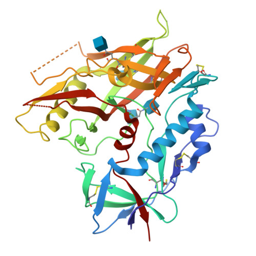
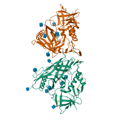
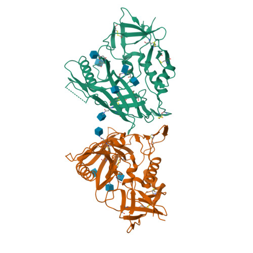
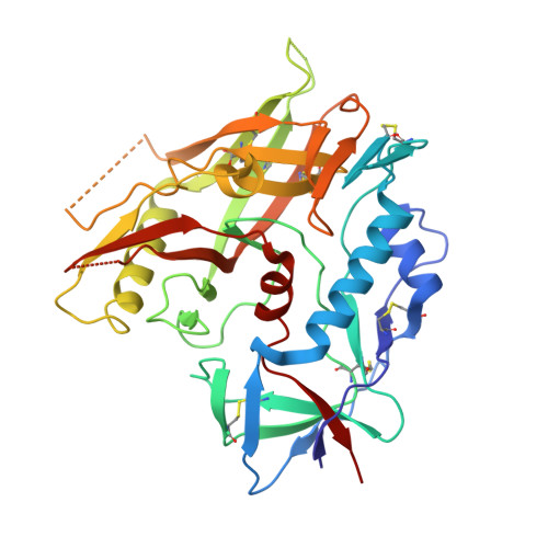
Unliganded HIV-1 gp120 core structures assume the CD4-bound conformation with regulation by quaternary interactions and variable loops.

Kwon, Y.D.Finzi, A.Wu, X.Dogo-Isonagie, C.Lee, L.K.Moore, L.R.Schmidt, S.D.Stuckey, J.Yang, Y.Zhou, T.Zhu, J.Vicic, D.A.Debnath, A.K.Shapiro, L.Bewley, C.A.Mascola, J.R.Sodroski, J.G.Kwong, P.D.

(2012) Proc Natl Acad Sci U S A 109: 5663-5668

  • DOI: https://doi.org/10.1073/pnas.1112391109
  • Primary Citation of Related Structures:  
    3TGQ, 3TGR, 3TGS, 3TGT, 3TIH

  • PubMed Abstract: 

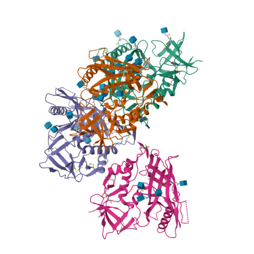
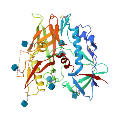
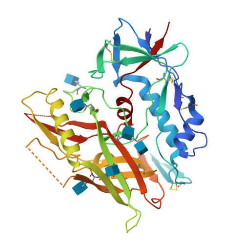
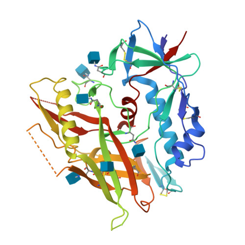
    The HIV-1 envelope (Env) spike (gp120(3)/gp41(3)) undergoes considerable structural rearrangements to mediate virus entry into cells and to evade the host immune response. Engagement of CD4, the primary human receptor, fixes a particular conformation and primes Env for entry. The CD4-bound state, however, is prone to spontaneous inactivation and susceptible to antibody neutralization. How does unliganded HIV-1 maintain CD4-binding capacity and regulate transitions to the CD4-bound state? To define this mechanistically, we determined crystal structures of unliganded core gp120 from HIV-1 clades B, C, and E. Notably, all of these unliganded HIV-1 structures resembled the CD4-bound state. Conformational fixation with ligand selection and thermodynamic analysis of full-length and core gp120 interactions revealed that the tendency of HIV-1 gp120 to adopt the CD4-bound conformation was restrained by the V1/V2- and V3-variable loops. In parallel, we determined the structure of core gp120 in complex with the small molecule, NBD-556, which specifically recognizes the CD4-bound conformation of gp120. Neutralization by NBD-556 indicated that Env spikes on primary isolates rarely assume the CD4-bound conformation spontaneously, although they could do so when quaternary restraints were loosened. Together, the results suggest that the CD4-bound conformation represents a "ground state" for the gp120 core, with variable loop and quaternary interactions restraining unliganded gp120 from "snapping" into this conformation. A mechanism of control involving deformations in unliganded structure from a functionally critical state (e.g., the CD4-bound state) provides advantages in terms of HIV-1 Env structural diversity and resistance to antibodies and inhibitors, while maintaining elements essential for entry.


  • Organizational Affiliation

    Vaccine Research Center, National Institute of Allergy and Infectious Diseases, National Institutes of Health, Bethesda, MD 20892, USA.


Macromolecules
Find similar proteins by:  (by identity cutoff)  |  3D Structure
Entity ID: 1
MoleculeChains Sequence LengthOrganismDetailsImage
HIV-1 YU2 gp120
A, B, C, D
352Human immunodeficiency virus 1Mutation(s): 0 
Gene Names: HIV-1 env
UniProt
Find proteins for P35961 (Human immunodeficiency virus type 1 group M subtype B (isolate YU-2))
Explore P35961 
Go to UniProtKB:  P35961
Entity Groups  
Sequence Clusters30% Identity50% Identity70% Identity90% Identity95% Identity100% Identity
UniProt GroupP35961
Glycosylation
Glycosylation Sites: 9
Sequence Annotations
Expand
  • Reference Sequence
Small Molecules
Ligands 1 Unique
IDChains Name / Formula / InChI Key2D Diagram3D Interactions
NAG
Query on NAG

Download Ideal Coordinates CCD File 
AA [auth D]
BA [auth D]
CA [auth D]
E [auth A]
F [auth A]
2-acetamido-2-deoxy-beta-D-glucopyranose
C8 H15 N O6
OVRNDRQMDRJTHS-FMDGEEDCSA-N
Experimental Data & Validation

Experimental Data

  • Method: X-RAY DIFFRACTION
  • Resolution: 3.40 Å
  • R-Value Free:  0.313 (Depositor), 0.300 (DCC) 
  • R-Value Work:  0.280 (Depositor), 0.270 (DCC) 
  • R-Value Observed: 0.281 (Depositor) 
Space Group: P 65
Unit Cell:
Length ( Å )Angle ( ˚ )
a = 222.97α = 90
b = 222.97β = 90
c = 86.48γ = 120
Software Package:
Software NamePurpose
PHENIXrefinement
PDB_EXTRACTdata extraction
HKL-2000data collection
HKL-2000data reduction
HKL-2000data scaling
PHASERphasing

Structure Validation

View Full Validation Report


Ligand Structure Quality Assessment 

Created with Raphaël 2.3.0Worse 01 BetterLigand structure goodness of fit to experimental dataBest fitted NAGClick on this verticalbar to view details

Entry History 

Deposition Data

Revision History  (Full details and data files)

  • Version 1.0: 2012-04-04
    Type: Initial release
  • Version 1.1: 2012-05-23
    Changes: Database references
  • Version 1.2: 2014-04-02
    Changes: Source and taxonomy
  • Version 1.3: 2020-07-29
    Type: Remediation
    Reason: Carbohydrate remediation
    Changes: Advisory, Data collection, Derived calculations, Structure summary
  • Version 1.4: 2023-09-13
    Changes: Advisory, Data collection, Database references, Refinement description, Structure summary
  • Version 1.5: 2024-10-16
    Changes: Structure summary