3RDC

Human Cyclophilin D Complexed with an Inhibitor


Experimental Data Snapshot

  • Method: X-RAY DIFFRACTION
  • Resolution: 1.94 Å
  • R-Value Free: 
    0.270 (Depositor), 0.270 (DCC) 
  • R-Value Work: 
    0.205 (Depositor), 0.210 (DCC) 
  • R-Value Observed: 
    0.208 (Depositor) 

Starting Model: experimental
View more details

wwPDB Validation   3D Report Full Report


Ligand Structure Quality Assessment 

Created with Raphaël 2.3.0Worse 01 BetterLigand structure goodness of fit to experimental dataBest fitted EA4Click on this verticalbar to view details

This is version 1.3 of the entry. See complete history


Literature

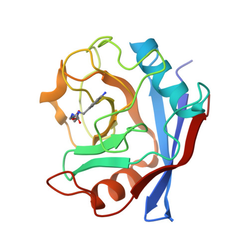
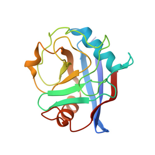
Combining 'dry' co-crystallization and in situ diffraction to facilitate ligand screening by X-ray crystallography.

Gelin, M.Delfosse, V.Allemand, F.Hoh, F.Sallaz-Damaz, Y.Pirocchi, M.Bourguet, W.Ferrer, J.L.Labesse, G.Guichou, J.F.

(2015) Acta Crystallogr D Biol Crystallogr 71: 1777-1787

  • DOI: https://doi.org/10.1107/S1399004715010342
  • Primary Citation of Related Structures:  
    3RDC, 4XLD, 4XN6, 4XNC, 4XNE, 4XOY, 4XOZ, 4XP0, 4XP2, 4XP3, 4XRJ, 4XRL, 4ZSC, 4ZSD

  • PubMed Abstract: 

    X-ray crystallography is an established technique for ligand screening in fragment-based drug-design projects, but the required manual handling steps - soaking crystals with ligand and the subsequent harvesting - are tedious and limit the throughput of the process. Here, an alternative approach is reported: crystallization plates are pre-coated with potential binders prior to protein crystallization and X-ray diffraction is performed directly 'in situ' (or in-plate). Its performance is demonstrated on distinct and relevant therapeutic targets currently being studied for ligand screening by X-ray crystallography using either a bending-magnet beamline or a rotating-anode generator. The possibility of using DMSO stock solutions of the ligands to be coated opens up a route to screening most chemical libraries.


  • Organizational Affiliation

    CNRS, UMR5048 - Université de Montpellier, Centre de Biochimie Structurale, 34090 Montpellier, France.


Macromolecules
Find similar proteins by:  (by identity cutoff)  |  3D Structure
Entity ID: 1
MoleculeChains Sequence LengthOrganismDetailsImage
Peptidyl-prolyl cis-trans isomerase F, mitochondrial166Homo sapiensMutation(s): 1 
Gene Names: PPIFCYP3
EC: 5.2.1.8
UniProt & NIH Common Fund Data Resources
Find proteins for P30405 (Homo sapiens)
Explore P30405 
Go to UniProtKB:  P30405
PHAROS:  P30405
GTEx:  ENSG00000108179 
Entity Groups  
Sequence Clusters30% Identity50% Identity70% Identity90% Identity95% Identity100% Identity
UniProt GroupP30405
Sequence Annotations
Expand
  • Reference Sequence
Small Molecules
Ligands 1 Unique
IDChains Name / Formula / InChI Key2D Diagram3D Interactions
EA4
Query on EA4

Download Ideal Coordinates CCD File 
B [auth A]ethyl N-[(4-aminobenzyl)carbamoyl]glycinate
C12 H17 N3 O3
UCMPTJMHPJQEED-UHFFFAOYSA-N
Experimental Data & Validation

Experimental Data

  • Method: X-RAY DIFFRACTION
  • Resolution: 1.94 Å
  • R-Value Free:  0.270 (Depositor), 0.270 (DCC) 
  • R-Value Work:  0.205 (Depositor), 0.210 (DCC) 
  • R-Value Observed: 0.208 (Depositor) 
Space Group: P 41 21 2
Unit Cell:
Length ( Å )Angle ( ˚ )
a = 56.445α = 90
b = 56.445β = 90
c = 86.995γ = 90
Software Package:
Software NamePurpose
MAR345dtbdata collection
X-PLORmodel building
REFMACrefinement
MOSFLMdata reduction
SCALAdata scaling
X-PLORphasing

Structure Validation

View Full Validation Report


Ligand Structure Quality Assessment 

Created with Raphaël 2.3.0Worse 01 BetterLigand structure goodness of fit to experimental dataBest fitted EA4Click on this verticalbar to view details

Entry History 

Deposition Data

Revision History  (Full details and data files)

  • Version 1.0: 2012-03-21
    Type: Initial release
  • Version 1.1: 2015-08-12
    Changes: Database references
  • Version 1.2: 2016-10-19
    Changes: Database references
  • Version 1.3: 2023-09-13
    Changes: Data collection, Database references, Derived calculations, Refinement description