3LN1

Structure of celecoxib bound at the COX-2 active site


Experimental Data Snapshot

  • Method: X-RAY DIFFRACTION
  • Resolution: 2.40 Å
  • R-Value Free: 
    0.264 (Depositor), 0.271 (DCC) 
  • R-Value Work: 
    0.232 (Depositor), 0.240 (DCC) 
  • R-Value Observed: 
    0.235 (Depositor) 

wwPDB Validation   3D Report Full Report


Ligand Structure Quality Assessment 

Created with Raphaël 2.3.0Worse 01 BetterLigand structure goodness of fit to experimental dataBest fitted HEMClick on this verticalbar to view detailsBest fitted CELClick on this verticalbar to view detailsBest fitted BOGClick on this verticalbar to view detailsBest fitted NAGClick on this verticalbar to view details

This is version 2.1 of the entry. See complete history


Literature

The novel benzopyran class of selective cyclooxygenase-2 inhibitors. Part 2: The second clinical candidate having a shorter and favorable human half-life.

Wang, J.L.Limburg, D.Graneto, M.J.Springer, J.Hamper, J.R.Liao, S.Pawlitz, J.L.Kurumbail, R.G.Maziasz, T.Talley, J.J.Kiefer, J.R.Carter, J.

(2010) Bioorg Med Chem Lett 20: 7159-7163

  • DOI: https://doi.org/10.1016/j.bmcl.2010.07.054
  • Primary Citation of Related Structures:  
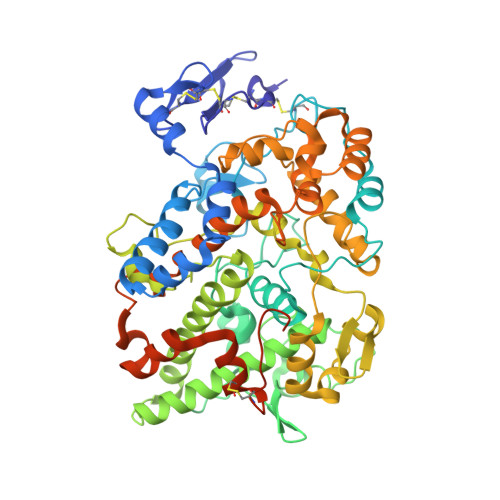
    3LN0, 3LN1, 3MQE, 3NTG

  • PubMed Abstract: 

    In this Letter, we provide the structure-activity relationships, optimization of design, testing criteria, and human half-life data for a series of selective COX-2 inhibitors. During the course of our structure-based drug design efforts, we discovered two distinct binding modes within the COX-2 active site for differently substituted members of this class. The challenge of a undesirably long human half-life for the first clinical candidate 1t(1/2)=360 h was addressed by multiple strategies, leading to the discovery of 29b-(S) (SC-75416) with t(1/2)=34 h.


  • Organizational Affiliation

    Pfizer Global Research and Development, Chesterfield, MO 63017, USA. jl47wang@yahoo.com


Macromolecules
Find similar proteins by:  (by identity cutoff)  |  3D Structure
Entity ID: 1
MoleculeChains Sequence LengthOrganismDetailsImage
Prostaglandin G/H synthase 2
A, B, C, D
587Mus musculusMutation(s): 0 
Gene Names: Cox-2Cox2Pghs-bprostaglandin H2 synthase 2Ptgs2Tis10
EC: 1.14.99.1
UniProt
Find proteins for Q05769 (Mus musculus)
Explore Q05769 
Go to UniProtKB:  Q05769
Entity Groups  
Sequence Clusters30% Identity50% Identity70% Identity90% Identity95% Identity100% Identity
UniProt GroupQ05769
Glycosylation
Glycosylation Sites: 3Go to GlyGen: Q05769-1
Sequence Annotations
Expand
  • Reference Sequence
Oligosaccharides

Help

Entity ID: 2
MoleculeChains Length2D Diagram Glycosylation3D Interactions
2-acetamido-2-deoxy-beta-D-glucopyranose-(1-4)-2-acetamido-2-deoxy-beta-D-glucopyranose-(1-4)-2-acetamido-2-deoxy-beta-D-glucopyranose
E, F, G, H
3N-Glycosylation
Glycosylation Resources
GlyTouCan:  G47362BJ
GlyCosmos:  G47362BJ
GlyGen:  G47362BJ
Small Molecules
Ligands 4 Unique
IDChains Name / Formula / InChI Key2D Diagram3D Interactions
HEM
Query on HEM

Download Ideal Coordinates CCD File 
K [auth A],
P [auth B],
T [auth C],
X [auth D]
PROTOPORPHYRIN IX CONTAINING FE
C34 H32 Fe N4 O4
KABFMIBPWCXCRK-RGGAHWMASA-L
CEL
Query on CEL

Download Ideal Coordinates CCD File 
L [auth A],
Q [auth B],
U [auth C],
Y [auth D]
4-[5-(4-METHYLPHENYL)-3-(TRIFLUOROMETHYL)-1H-PYRAZOL-1-YL]BENZENESULFONAMIDE
C17 H14 F3 N3 O2 S
RZEKVGVHFLEQIL-UHFFFAOYSA-N
BOG
Query on BOG

Download Ideal Coordinates CCD File 
M [auth A],
Z [auth D]
octyl beta-D-glucopyranoside
C14 H28 O6
HEGSGKPQLMEBJL-RKQHYHRCSA-N
NAG
Query on NAG

Download Ideal Coordinates CCD File 
I [auth A]
J [auth A]
N [auth B]
O [auth B]
R [auth C]
2-acetamido-2-deoxy-beta-D-glucopyranose
C8 H15 N O6
OVRNDRQMDRJTHS-FMDGEEDCSA-N
Experimental Data & Validation

Experimental Data

  • Method: X-RAY DIFFRACTION
  • Resolution: 2.40 Å
  • R-Value Free:  0.264 (Depositor), 0.271 (DCC) 
  • R-Value Work:  0.232 (Depositor), 0.240 (DCC) 
  • R-Value Observed: 0.235 (Depositor) 
Space Group: P 21 21 2
Unit Cell:
Length ( Å )Angle ( ˚ )
a = 180.942α = 90
b = 135.377β = 90
c = 124.079γ = 90
Software Package:
Software NamePurpose
MAR345dtbdata collection
PHASERphasing
REFMACrefinement
HKL-2000data reduction
HKL-2000data scaling

Structure Validation

View Full Validation Report


Ligand Structure Quality Assessment 

Created with Raphaël 2.3.0Worse 01 BetterLigand structure goodness of fit to experimental dataBest fitted HEMClick on this verticalbar to view detailsBest fitted CELClick on this verticalbar to view detailsBest fitted BOGClick on this verticalbar to view detailsBest fitted NAGClick on this verticalbar to view details

Entry History 

Deposition Data

Revision History  (Full details and data files)

  • Version 1.0: 2010-10-27
    Type: Initial release
  • Version 1.1: 2011-07-13
    Changes: Version format compliance
  • Version 2.0: 2020-07-29
    Type: Remediation
    Reason: Carbohydrate remediation
    Changes: Atomic model, Data collection, Derived calculations, Structure summary
  • Version 2.1: 2024-10-16
    Changes: Data collection, Database references, Refinement description, Structure summary