3KFS

HIV Protease (PR) dimer with inhibitor TL-3 bound and fragment 2F4 in the outside/top of flap


Experimental Data Snapshot

  • Method: X-RAY DIFFRACTION
  • Resolution: 1.80 Å
  • R-Value Free: 0.250 
  • R-Value Work: 0.203 
  • R-Value Observed: 0.205 

Starting Model: experimental
View more details

wwPDB Validation   3D Report Full Report


Ligand Structure Quality Assessment 

Created with Raphaël 2.3.0Worse 01 BetterLigand structure goodness of fit to experimental dataBest fitted 3TLClick on this verticalbar to view detailsBest fitted 2FXClick on this verticalbar to view details

This is version 1.4 of the entry. See complete history


Literature

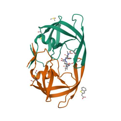
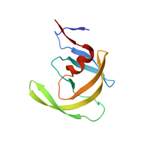
Fragment-based screen against HIV protease.

Perryman, A.L.Zhang, Q.Soutter, H.H.Rosenfeld, R.McRee, D.E.Olson, A.J.Elder, J.E.David Stout, C.

(2010) Chem Biol Drug Des 75: 257-268

  • DOI: https://doi.org/10.1111/j.1747-0285.2009.00943.x
  • Primary Citation of Related Structures:  
    3KF0, 3KFN, 3KFP, 3KFR, 3KFS, 4E43

  • PubMed Abstract: 

    We have employed a fragment-based screen against wild-type (NL4-3) HIV protease (PR) using the Active Sight fragment library and X-ray crystallography. The experiments reveal two new binding sites for small molecules. PR was co-crystallized with fragments, or crystals were soaked in fragment solutions, using five crystal forms, and 378 data sets were collected to 2.3-1.3 A resolution. Fragment binding induces a distinct conformation and specific crystal form of TL-3 inhibited PR during co-crystallization. One fragment, 2-methylcyclohexanol, binds in the 'exo site' adjacent to the Gly(16)Gly(17)Gln(18)loop where the amide of Gly(17)is a specific hydrogen bond donor, and hydrophobic contacts occur with the side chains of Lys(14)and Leu(63). Another fragment, indole-6-carboxylic acid, binds on the 'outside/top of the flap' via hydrophobic contacts with Trp(42), Pro(44), Met(46), and Lys(55), a hydrogen bond with Val(56), and a salt-bridge with Arg(57). 2-acetyl-benzothiophene also binds at this site. This study is the first fragment-based crystallographic screen against HIV PR, and the first time that fragments were screened against an inhibitor-bound drug target to search for compounds that both bind to novel sites and stabilize the inhibited conformation of the target.


  • Organizational Affiliation

    Department of Molecular Biology, The Scripps Research Institute, 10550 N. Torrey Pines Rd., La Jolla, CA 92037, USA.


Macromolecules
Find similar proteins by:  (by identity cutoff)  |  3D Structure
Entity ID: 1
MoleculeChains Sequence LengthOrganismDetailsImage
Protease
A, B
99Human immunodeficiency virus 1Mutation(s): 1 
Gene Names: POL
EC: 3.4.23.16
UniProt
Find proteins for P12499 (Human immunodeficiency virus type 1 group M subtype D (isolate Z2/CDC-Z34))
Explore P12499 
Go to UniProtKB:  P12499
Entity Groups  
Sequence Clusters30% Identity50% Identity70% Identity90% Identity95% Identity100% Identity
UniProt GroupP12499
Sequence Annotations
Expand
  • Reference Sequence
Small Molecules
Ligands 4 Unique
IDChains Name / Formula / InChI Key2D Diagram3D Interactions
3TL
Query on 3TL

Download Ideal Coordinates CCD File 
C [auth A]benzyl [(1S,4S,7S,8R,9R,10S,13S,16S)-7,10-dibenzyl-8,9-dihydroxy-1,16-dimethyl-4,13-bis(1-methylethyl)-2,5,12,15,18-pentaoxo-20-phenyl-19-oxa-3,6,11,14,17-pentaazaicos-1-yl]carbamate
C50 H64 N6 O10
BJJPNOGMLLUCER-KUTQPOQPSA-N
2FX
Query on 2FX

Download Ideal Coordinates CCD File 
G [auth B]1-benzothiophen-2-ylacetic acid
C10 H8 O2 S
SAAWNQLNUJBOLW-UHFFFAOYSA-N
BME
Query on BME

Download Ideal Coordinates CCD File 
F [auth A],
I [auth B]
BETA-MERCAPTOETHANOL
C2 H6 O S
DGVVWUTYPXICAM-UHFFFAOYSA-N
DMS
Query on DMS

Download Ideal Coordinates CCD File 
D [auth A],
E [auth A],
H [auth B]
DIMETHYL SULFOXIDE
C2 H6 O S
IAZDPXIOMUYVGZ-UHFFFAOYSA-N
Binding Affinity Annotations 
IDSourceBinding Affinity
3TL BindingDB:  3KFS Ki: 1.5 (nM) from 1 assay(s)
Biologically Interesting Molecules (External Reference) 1 Unique
Experimental Data & Validation

Experimental Data

  • Method: X-RAY DIFFRACTION
  • Resolution: 1.80 Å
  • R-Value Free: 0.250 
  • R-Value Work: 0.203 
  • R-Value Observed: 0.205 
  • Space Group: P 21 21 21
Unit Cell:
Length ( Å )Angle ( ˚ )
a = 28.95α = 90
b = 66.35β = 90
c = 92.97γ = 90
Software Package:
Software NamePurpose
REFMACrefinement
PDB_EXTRACTdata extraction
Blu-Icedata collection
MOSFLMdata reduction
SCALAdata scaling
MOLREPphasing

Structure Validation

View Full Validation Report


Ligand Structure Quality Assessment 

Created with Raphaël 2.3.0Worse 01 BetterLigand structure goodness of fit to experimental dataBest fitted 3TLClick on this verticalbar to view detailsBest fitted 2FXClick on this verticalbar to view details

Entry History 

Deposition Data

  • Released Date: 2010-02-23 
  • Deposition Author(s): Stout, C.D.

Revision History  (Full details and data files)

  • Version 1.0: 2010-02-23
    Type: Initial release
  • Version 1.1: 2011-07-13
    Changes: Atomic model, Database references, Derived calculations, Non-polymer description, Structure summary, Version format compliance
  • Version 1.2: 2013-02-27
    Changes: Other
  • Version 1.3: 2021-10-13
    Changes: Database references, Derived calculations
  • Version 1.4: 2023-09-06
    Changes: Data collection, Refinement description