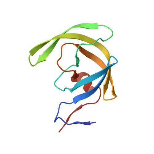
3KDC

Crystal Structure of HIV-1 Protease (Q7K, L33I, L63I) in Complex with KNI-10074


Experimental Data Snapshot

  • Method: X-RAY DIFFRACTION
  • Resolution: 2.20 Å
  • R-Value Free: 0.237 
  • R-Value Work: 0.177 
  • R-Value Observed: 0.180 

Starting Model: experimental
View more details

wwPDB Validation   3D Report Full Report


Ligand Structure Quality Assessment 


This is version 1.4 of the entry. See complete history


Literature

How much binding affinity can be gained by filling a cavity?

Kawasaki, Y.Chufan, E.E.Lafont, V.Hidaka, K.Kiso, Y.Mario Amzel, L.Freire, E.

(2010) Chem Biol Drug Des 75: 143-151

  • DOI: https://doi.org/10.1111/j.1747-0285.2009.00921.x
  • Primary Citation of Related Structures:  
    3KDB, 3KDC, 3KDD

  • PubMed Abstract: 

    Binding affinity optimization is critical during drug development. Here, we evaluate the thermodynamic consequences of filling a binding cavity with functionalities of increasing van der Waals radii (-H, -F, -Cl, and CH(3)) that improve the geometric fit without participating in hydrogen bonding or other specific interactions. We observe a binding affinity increase of two orders of magnitude. There appears to be three phases in the process. The first phase is associated with the formation of stable van der Waals interactions. This phase is characterized by a gain in binding enthalpy and a loss in binding entropy, attributed to a loss of conformational degrees of freedom. For the specific case presented in this article, the enthalpy gain amounts to -1.5 kcal/mol while the entropic losses amount to +0.9 kcal/mol resulting in a net 3.5-fold affinity gain. The second phase is characterized by simultaneous enthalpic and entropic gains. This phase improves the binding affinity 25-fold. The third phase represents the collapse of the trend and is triggered by the introduction of chemical functionalities larger than the binding cavity itself [CH(CH(3))(2)]. It is characterized by large enthalpy and affinity losses. The thermodynamic signatures associated with each phase provide guidelines for lead optimization.


  • Organizational Affiliation

    Department of Biology, Johns Hopkins University, Baltimore, MD 21218, USA.


Macromolecules
Find similar proteins by:  (by identity cutoff)  |  3D Structure
Entity ID: 1
MoleculeChains Sequence LengthOrganismDetailsImage
Protease
A, B
99Human immunodeficiency virus type 1 (BRU ISOLATE)Mutation(s): 3 
Gene Names: gag-pol
EC: 3.4.23.16
UniProt
Find proteins for P03367 (Human immunodeficiency virus type 1 group M subtype B (isolate BRU/LAI))
Explore P03367 
Go to UniProtKB:  P03367
Entity Groups  
Sequence Clusters30% Identity50% Identity70% Identity90% Identity95% Identity100% Identity
UniProt GroupP03367
Sequence Annotations
Expand
  • Reference Sequence
Small Molecules
Ligands 3 Unique
IDChains Name / Formula / InChI Key2D Diagram3D Interactions
JZP
Query on JZP

Download Ideal Coordinates CCD File 
C [auth A](4R)-3-[(2S,3S)-3-{[(2,6-dichlorophenoxy)acetyl]amino}-2-hydroxy-4-phenylbutanoyl]-N-[(1S,2R)-2-hydroxy-2,3-dihydro-1H-inden-1-yl]-5,5-dimethyl-1,3-thiazolidine-4-carboxamide
C33 H35 Cl2 N3 O6 S
XMAMZSZKRBFNLW-UPZHSWQNSA-N
GOL
Query on GOL

Download Ideal Coordinates CCD File 
D [auth A]GLYCEROL
C3 H8 O3
PEDCQBHIVMGVHV-UHFFFAOYSA-N
CL
Query on CL

Download Ideal Coordinates CCD File 
E [auth A],
F [auth A],
G [auth B],
H [auth B]
CHLORIDE ION
Cl
VEXZGXHMUGYJMC-UHFFFAOYSA-M
Binding Affinity Annotations 
IDSourceBinding Affinity
JZP PDBBind:  3KDC Kd: 0.52 (nM) from 1 assay(s)
Biologically Interesting Molecules (External Reference) 1 Unique
Experimental Data & Validation

Experimental Data

  • Method: X-RAY DIFFRACTION
  • Resolution: 2.20 Å
  • R-Value Free: 0.237 
  • R-Value Work: 0.177 
  • R-Value Observed: 0.180 
  • Space Group: P 21 21 2
Unit Cell:
Length ( Å )Angle ( ˚ )
a = 58.31α = 90
b = 86.095β = 90
c = 46.369γ = 90
Software Package:
Software NamePurpose
HKL-2000data collection
AMoREphasing
REFMACrefinement
HKL-2000data reduction
HKL-2000data scaling

Structure Validation

View Full Validation Report


Ligand Structure Quality Assessment 


Entry History 

Deposition Data

Revision History  (Full details and data files)

  • Version 1.0: 2010-03-16
    Type: Initial release
  • Version 1.1: 2011-07-13
    Changes: Atomic model, Database references, Derived calculations, Non-polymer description, Structure summary, Version format compliance
  • Version 1.2: 2012-12-12
    Changes: Other
  • Version 1.3: 2021-10-13
    Changes: Database references, Derived calculations
  • Version 1.4: 2023-09-06
    Changes: Data collection, Refinement description