3GD7 | pdb_00003gd7

Crystal structure of human NBD2 complexed with N6-Phenylethyl-ATP (P-ATP)


Experimental Data Snapshot

  • Method: X-RAY DIFFRACTION
  • Resolution: 2.70 Å
  • R-Value Free: 
    0.308 (Depositor), 0.300 (DCC) 
  • R-Value Work: 
    0.247 (Depositor), 0.250 (DCC) 

Starting Model: experimental
View more details

wwPDB Validation   3D Report Full Report


Ligand Structure Quality Assessment 

Created with Raphaël 2.3.0Worse 01 BetterLigand structure goodness of fit to experimental dataBest fitted B44Click on this verticalbar to view details

This is version 1.5 of the entry. See complete history


Literature

Crystal structure of human NBD2 complexed with N6-Phenylethyl-ATP (P-ATP)

Atwell, S.Antonysamy, S.Guggino, W.B.Conners, K.Emtage, S.Gheyi, T.Hunt, J.F.Lewis, H.A.Lu, F.Sauder, J.M.Weber, P.C.Wetmore, D.Zhao, X.

To be published.

Macromolecules
Find similar proteins by:  (by identity cutoff)  |  3D Structure
Entity ID: 1
MoleculeChains Sequence LengthOrganismDetailsImage
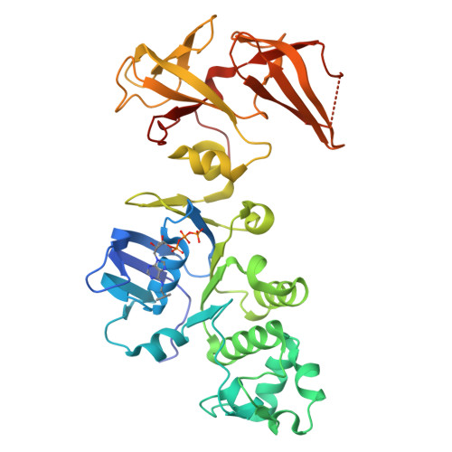
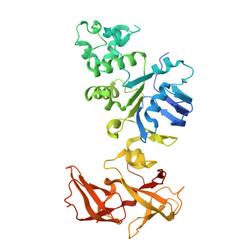
Fusion complex of Cystic fibrosis transmembrane conductance regulator, residues 1193-1427 and Maltose/maltodextrin import ATP-binding protein malK, residues 219-371
A, B, C, D
390Homo sapiensEscherichia coli K-12
This entity is chimeric
Mutation(s): 7 
Gene Names: CFTRmalK
EC: 5.6.1.6 (UniProt), 7.5.2.1 (UniProt)
UniProt & NIH Common Fund Data Resources
Find proteins for P13569 (Homo sapiens)
Explore P13569 
Go to UniProtKB:  P13569
PHAROS:  P13569
GTEx:  ENSG00000001626 
Find proteins for P68187 (Escherichia coli (strain K12))
Explore P68187 
Go to UniProtKB:  P68187
Entity Groups  
Sequence Clusters30% Identity50% Identity70% Identity90% Identity95% Identity100% Identity
UniProt GroupsP13569P68187
Sequence Annotations
Expand
  • Reference Sequence
Experimental Data & Validation

Experimental Data

  • Method: X-RAY DIFFRACTION
  • Resolution: 2.70 Å
  • R-Value Free:  0.308 (Depositor), 0.300 (DCC) 
  • R-Value Work:  0.247 (Depositor), 0.250 (DCC) 
Space Group: P 21 21 2
Unit Cell:
Length ( Å )Angle ( ˚ )
a = 137.837α = 90
b = 173.725β = 90
c = 82.574γ = 90
Software Package:
Software NamePurpose
REFMACrefinement
PDB_EXTRACTdata extraction
ADSCdata collection
d*TREKdata reduction
SCALAdata scaling
PHASERphasing

Structure Validation

View Full Validation Report


Ligand Structure Quality Assessment 

Created with Raphaël 2.3.0Worse 01 BetterLigand structure goodness of fit to experimental dataBest fitted B44Click on this verticalbar to view details

Entry History 

Deposition Data

Revision History  (Full details and data files)

  • Version 1.0: 2010-03-02
    Type: Initial release
  • Version 1.1: 2011-07-13
    Changes: Version format compliance
  • Version 1.2: 2017-08-16
    Changes: Refinement description, Source and taxonomy
  • Version 1.3: 2018-11-21
    Changes: Data collection, Structure summary
  • Version 1.4: 2021-10-20
    Changes: Database references, Derived calculations
  • Version 1.5: 2023-09-06
    Changes: Data collection, Refinement description