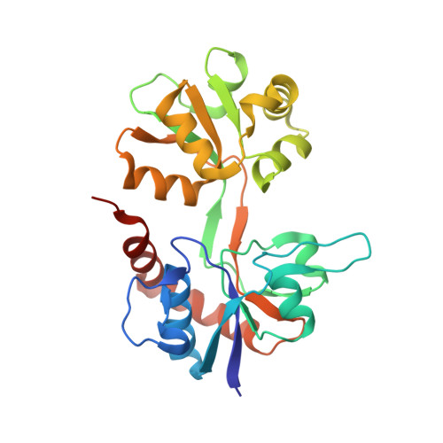
2QS3 | pdb_00002qs3

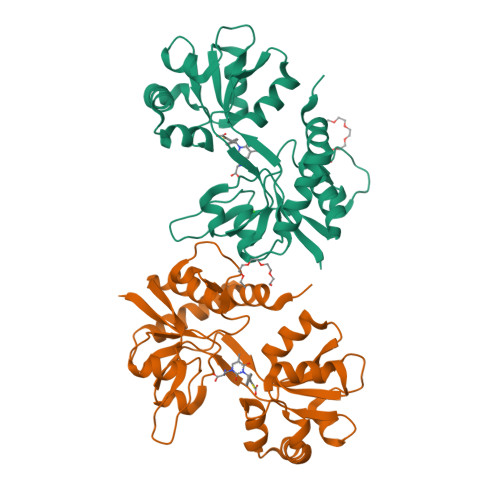
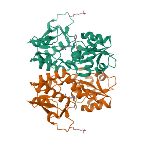
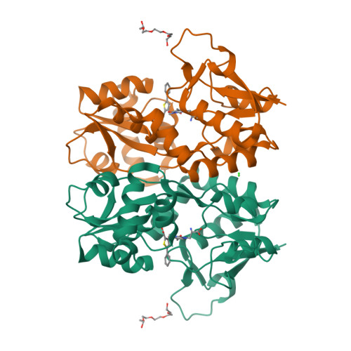
Crystal structure of the GluR5 ligand binding core dimer in complex with UBP316 at 1.76 Angstroms resolution


Experimental Data Snapshot

  • Method: X-RAY DIFFRACTION
  • Resolution: 1.76 Å
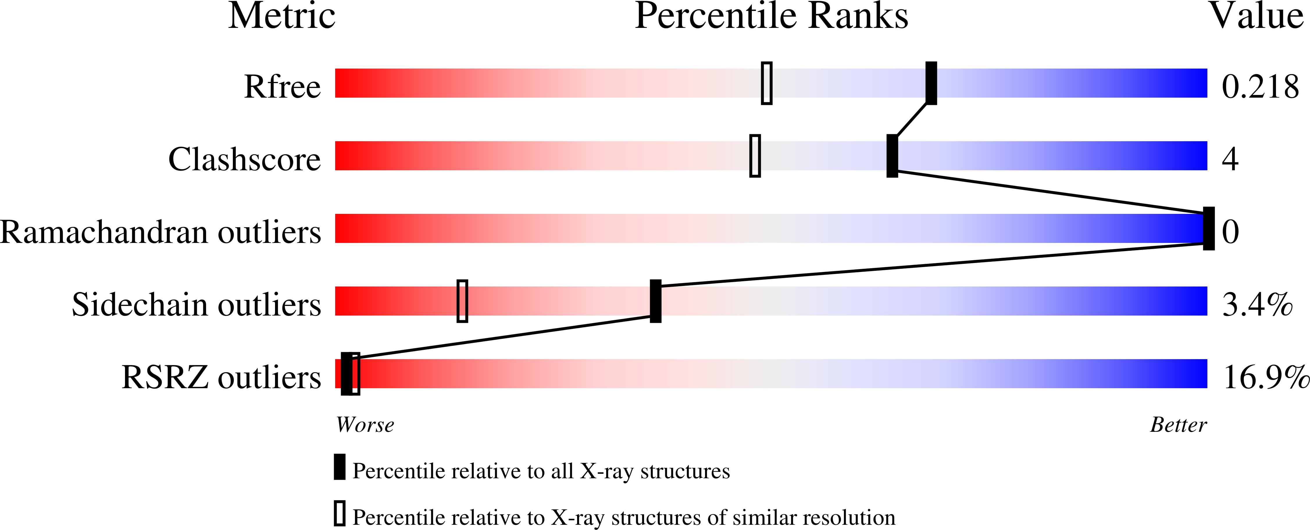
  • R-Value Free: 
    0.218 (Depositor), 0.220 (DCC) 
  • R-Value Work: 
    0.187 (Depositor), 0.190 (DCC) 
  • R-Value Observed: 
    0.189 (Depositor) 

Starting Model: experimental
View more details

wwPDB Validation   3D Report Full Report


Ligand Structure Quality Assessment 

Created with Raphaël 2.3.0Worse 01 BetterLigand structure goodness of fit to experimental dataBest fitted UBEClick on this verticalbar to view details

This is version 1.5 of the entry. See complete history


Literature

ACET is a highly potent and specific kainate receptor antagonist: characterisation and effects on hippocampal mossy fibre function.

Dargan, S.L.Clarke, V.R.Alushin, G.M.Sherwood, J.L.Nistico, R.Bortolotto, Z.A.Ogden, A.M.Bleakman, D.Doherty, A.J.Lodge, D.Mayer, M.L.Fitzjohn, S.M.Jane, D.E.Collingridge, G.L.

(2009) Neuropharmacology 56: 121-130

  • DOI: https://doi.org/10.1016/j.neuropharm.2008.08.016
  • Primary Citation of Related Structures:  
    2QS3

  • PubMed Abstract: 

    Kainate receptors (KARs) are involved in both NMDA receptor-independent long-term potentiation (LTP) and synaptic facilitation at mossy fibre synapses in the CA3 region of the hippocampus. However, the identity of the KAR subtypes involved remains controversial. Here we used a highly potent and selective GluK1 (formerly GluR5) antagonist (ACET) to elucidate roles of GluK1-containing KARs in these synaptic processes. We confirmed that ACET is an extremely potent GluK1 antagonist, with a Kb value of 1.4+/-0.2 nM. In contrast, ACET was ineffective at GluK2 (formerly GluR6) receptors at all concentrations tested (up to 100 microM) and had no effect at GluK3 (formerly GluR7) when tested at 1 microM. The X-ray crystal structure of ACET bound to the ligand binding core of GluK1 was similar to the UBP310-GluK1 complex. In the CA1 region of hippocampal slices, ACET was effective at blocking the depression of both fEPSPs and monosynaptically evoked GABAergic transmission induced by ATPA, a GluK1 selective agonist. In the CA3 region of the hippocampus, ACET blocked the induction of NMDA receptor-independent mossy fibre LTP. To directly investigate the role of pre-synaptic GluK1-containing KARs we combined patch-clamp electrophysiology and 2-photon microscopy to image Ca2+ dynamics in individual giant mossy fibre boutons. ACET consistently reduced short-term facilitation of pre-synaptic calcium transients induced by 5 action potentials evoked at 20-25Hz. Taken together our data provide further evidence for a physiological role of GluK1-containing KARs in synaptic facilitation and LTP induction at mossy fibre-CA3 synapses.


  • Organizational Affiliation

    MRC Centre for Synaptic Plasticity, University of Bristol, Bristol BS8 1TD, UK. sheila.dargan@bristol.ac.uk


Macromolecules
Find similar proteins by:  (by identity cutoff)  |  3D Structure
Entity ID: 1
MoleculeChains Sequence LengthOrganismDetailsImage
Glutamate receptor, ionotropic kainate 1
A, B
258Rattus norvegicusMutation(s): 1 
Gene Names: Grik1Glur5
UniProt
Find proteins for P22756 (Rattus norvegicus)
Explore P22756 
Go to UniProtKB:  P22756
Entity Groups  
Sequence Clusters30% Identity50% Identity70% Identity90% Identity95% Identity100% Identity
UniProt GroupP22756
Sequence Annotations
Expand
  • Reference Sequence
Small Molecules
Ligands 3 Unique
IDChains Name / Formula / InChI Key2D Diagram3D Interactions
UBE
Query on UBE

Download Ideal Coordinates CCD File 
E [auth A],
H [auth B]
3-({3-[(2S)-2-amino-2-carboxyethyl]-5-methyl-2,6-dioxo-3,6-dihydropyrimidin-1(2H)-yl}methyl)-5-phenylthiophene-2-carboxylic acid
C20 H19 N3 O6 S
LCZDCKMQSBGXAH-AWEZNQCLSA-N
1PE
Query on 1PE

Download Ideal Coordinates CCD File 
D [auth A],
G [auth B]
PENTAETHYLENE GLYCOL
C10 H22 O6
JLFNLZLINWHATN-UHFFFAOYSA-N
CL
Query on CL

Download Ideal Coordinates CCD File 
C [auth A],
F [auth B]
CHLORIDE ION
Cl
VEXZGXHMUGYJMC-UHFFFAOYSA-M
Experimental Data & Validation

Experimental Data

  • Method: X-RAY DIFFRACTION
  • Resolution: 1.76 Å
  • R-Value Free:  0.218 (Depositor), 0.220 (DCC) 
  • R-Value Work:  0.187 (Depositor), 0.190 (DCC) 
  • R-Value Observed: 0.189 (Depositor) 
Space Group: C 2 2 21
Unit Cell:
Length ( Å )Angle ( ˚ )
a = 97.766α = 90
b = 97.702β = 90
c = 128.604γ = 90
Software Package:
Software NamePurpose
REFMACrefinement
SERGUIdata collection
HKL-2000data reduction
HKL-2000data scaling

Structure Validation

View Full Validation Report


Ligand Structure Quality Assessment 

Created with Raphaël 2.3.0Worse 01 BetterLigand structure goodness of fit to experimental dataBest fitted UBEClick on this verticalbar to view details

Entry History 

Deposition Data

Revision History  (Full details and data files)

  • Version 1.0: 2008-08-05
    Type: Initial release
  • Version 1.1: 2011-07-13
    Changes: Advisory, Version format compliance
  • Version 1.2: 2012-08-22
    Changes: Database references
  • Version 1.3: 2017-08-02
    Changes: Source and taxonomy
  • Version 1.4: 2021-10-20
    Changes: Database references, Derived calculations
  • Version 1.5: 2023-08-30
    Changes: Data collection, Refinement description