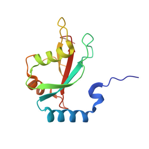
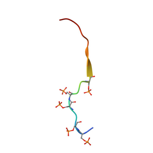
Structural basis for phosphorylation-triggered autophagic clearance of Salmonella.
Rogov, V.V., Suzuki, H., Fiskin, E., Wild, P., Kniss, A., Rozenknop, A., Kato, R., Kawasaki, M., McEwan, D.G., Lohr, F., Guntert, P., Dikic, I., Wakatsuki, S., Dotsch, V.(2013) Biochem J 454: 459-466
- PubMed: 23805866
- DOI: https://doi.org/10.1042/BJ20121907
- Primary Citation of Related Structures:
2LUE, 3VTU, 3VTV, 3VTW - PubMed Abstract:
Selective autophagy is mediated by the interaction of autophagy modifiers and autophagy receptors that also bind to ubiquitinated cargo. Optineurin is an autophagy receptor that plays a role in the clearance of cytosolic Salmonella. The interaction between receptors and modifiers is often relatively weak, with typical values for the dissociation constant in the low micromolar range. The interaction of optineurin with autophagy modifiers is even weaker, but can be significantly enhanced through phosphorylation by the TBK1 {TANK [TRAF (tumour-necrosis-factor-receptor-associated factor)-associated nuclear factor κB activator]-binding kinase 1}. In the present study we describe the NMR and crystal structures of the autophagy modifier LC3B (microtubule-associated protein light chain 3 beta) in complex with the LC3 interaction region of optineurin either phosphorylated or bearing phospho-mimicking mutations. The structures show that the negative charge induced by phosphorylation is recognized by the side chains of Arg¹¹ and Lys⁵¹ in LC3B. Further mutational analysis suggests that the replacement of the canonical tryptophan residue side chain of autophagy receptors with the smaller phenylalanine side chain in optineurin significantly weakens its interaction with the autophagy modifier LC3B. Through phosphorylation of serine residues directly N-terminally located to the phenylalanine residue, the affinity is increased to the level normally seen for receptor-modifier interactions. Phosphorylation, therefore, acts as a switch for optineurin-based selective autophagy.
Organizational Affiliation:
Institute of Biophysical Chemistry and Center for Biomolecular Magnetic Resonance, Goethe University, Max-van-Laue Str. 9, 60438 Frankfurt am Main, Germany.


