2J9P

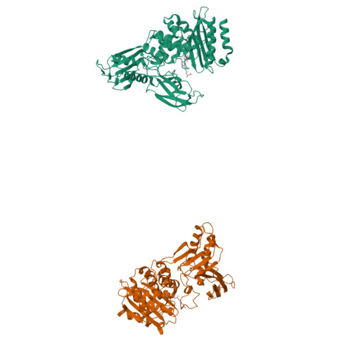
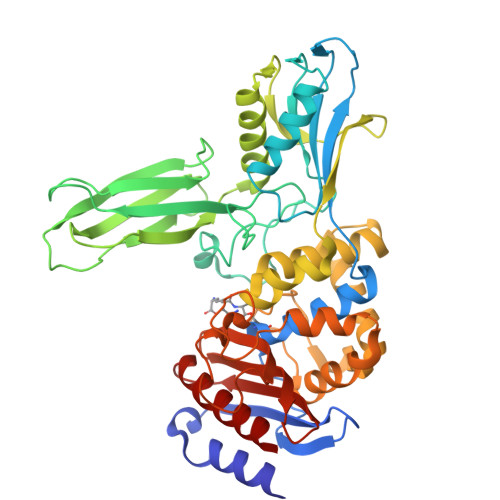
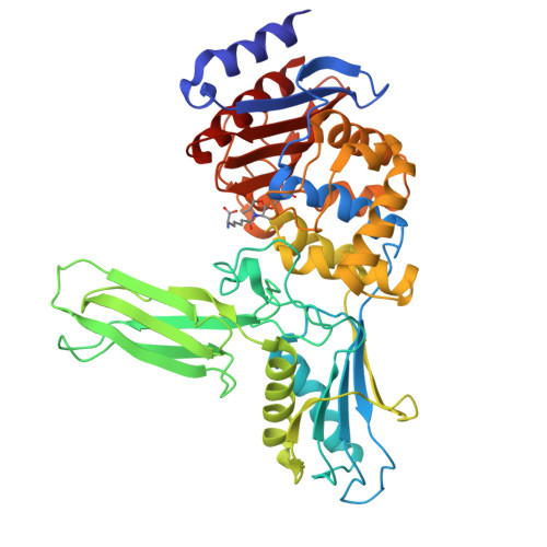
Crystal structure of the Bacillus subtilis PBP4a, and its complex with a peptidoglycan mimetic peptide.


Experimental Data Snapshot

  • Method: X-RAY DIFFRACTION
  • Resolution: 2.80 Å
  • R-Value Free: 0.290 
  • R-Value Work: 0.208 
  • R-Value Observed: 0.208 

Starting Model: experimental
View more details

wwPDB Validation   3D Report Full Report


Ligand Structure Quality Assessment 


This is version 1.5 of the entry. See complete history


Literature

Crystal structure of the Bacillus subtilis penicillin-binding protein 4a, and its complex with a peptidoglycan mimetic peptide.

Sauvage, E.Duez, C.Herman, R.Kerff, F.Petrella, S.Anderson, J.W.Adediran, S.A.Pratt, R.F.Frere, J.M.Charlier, P.

(2007) J Mol Biol 371: 528-539

  • DOI: https://doi.org/10.1016/j.jmb.2007.05.071
  • Primary Citation of Related Structures:  
    1W5D, 2J9P

  • PubMed Abstract: 

    The genome of Bacillus subtilis encodes 16 penicillin-binding proteins (PBPs) involved in the synthesis and/or remodelling of the peptidoglycan during the complex life cycle of this sporulating Gram-positive rod-shaped bacterium. PBP4a (encoded by the dacC gene) is a low-molecular mass PBP clearly exhibiting in vitro DD-carboxypeptidase activity. We have solved the crystal structure of this protein alone and in complex with a peptide (D-alpha-aminopymelyl-epsilon-D-alanyl-D-alanine) that mimics the C-terminal end of the Bacillus peptidoglycan stem peptide. PBP4a is composed of three domains: the penicillin-binding domain with a fold similar to the class A beta-lactamase structure and two domains inserted between the conserved motifs 1 and 2 characteristic of the penicillin-recognizing enzymes. The soaking of PBP4a in a solution of D-alpha-aminopymelyl-epsilon-D-alanyl-D-alanine resulted in an adduct between PBP4a and a D-alpha-aminopimelyl-epsilon-D-alanine dipeptide and an unbound D-alanine, i.e. the products of acylation of PBP4a by D-alpha-aminopymelyl-epsilon-D-alanyl-D-alanine with the release of a D-alanine. The adduct also reveals a binding pocket specific to the diaminopimelic acid, the third residue of the peptidoglycan stem pentapeptide of B. subtilis. This pocket is specific for this class of PBPs.


  • Organizational Affiliation

    Centre d'Ingénierie des Protéines, Université de Liège, Institut de Physique B5 et Institut de Chimie B6a, Sart Tilman, B-4000 Liège, Belgium. [email protected]


Macromolecules
Find similar proteins by:  (by identity cutoff)  |  3D Structure
Entity ID: 1
MoleculeChains Sequence LengthOrganismDetailsImage
D-alanyl-D-alanine carboxypeptidase DacC
A, B
462Bacillus subtilis subsp. subtilis str. 168Mutation(s): 0 
Gene Names: dacCpbpBSU18350
EC: 3.4.16.4
UniProt
Find proteins for P39844 (Bacillus subtilis (strain 168))
Explore P39844 
Go to UniProtKB:  P39844
Entity Groups  
Sequence Clusters30% Identity50% Identity70% Identity90% Identity95% Identity100% Identity
UniProt GroupP39844
Sequence Annotations
Expand
  • Reference Sequence
Experimental Data & Validation

Experimental Data

  • Method: X-RAY DIFFRACTION
  • Resolution: 2.80 Å
  • R-Value Free: 0.290 
  • R-Value Work: 0.208 
  • R-Value Observed: 0.208 
  • Space Group: P 21 21 21
Unit Cell:
Length ( Å )Angle ( ˚ )
a = 73.859α = 90
b = 77.586β = 90
c = 164.739γ = 90
Software Package:
Software NamePurpose
CNSrefinement
MOSFLMdata reduction
CCP4data scaling
AMoREphasing

Structure Validation

View Full Validation Report


Ligand Structure Quality Assessment 


Entry History 

Deposition Data

Revision History  (Full details and data files)

  • Version 1.0: 2007-07-03
    Type: Initial release
  • Version 1.1: 2011-05-08
    Changes: Version format compliance
  • Version 1.2: 2011-07-13
    Changes: Version format compliance
  • Version 1.3: 2018-12-12
    Changes: Data collection, Database references, Source and taxonomy, Structure summary
  • Version 1.4: 2023-12-13
    Changes: Data collection, Database references, Derived calculations, Other, Refinement description
  • Version 1.5: 2024-11-06
    Changes: Structure summary