2V2S

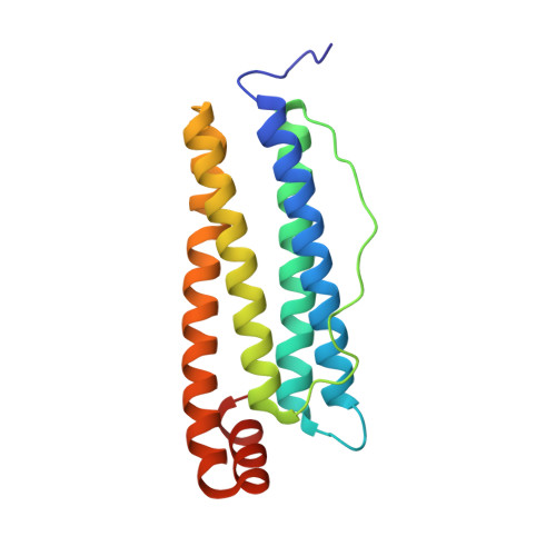
Mutant R59M recombinant horse spleen apoferritin crystallized in acidic conditions


Experimental Data Snapshot

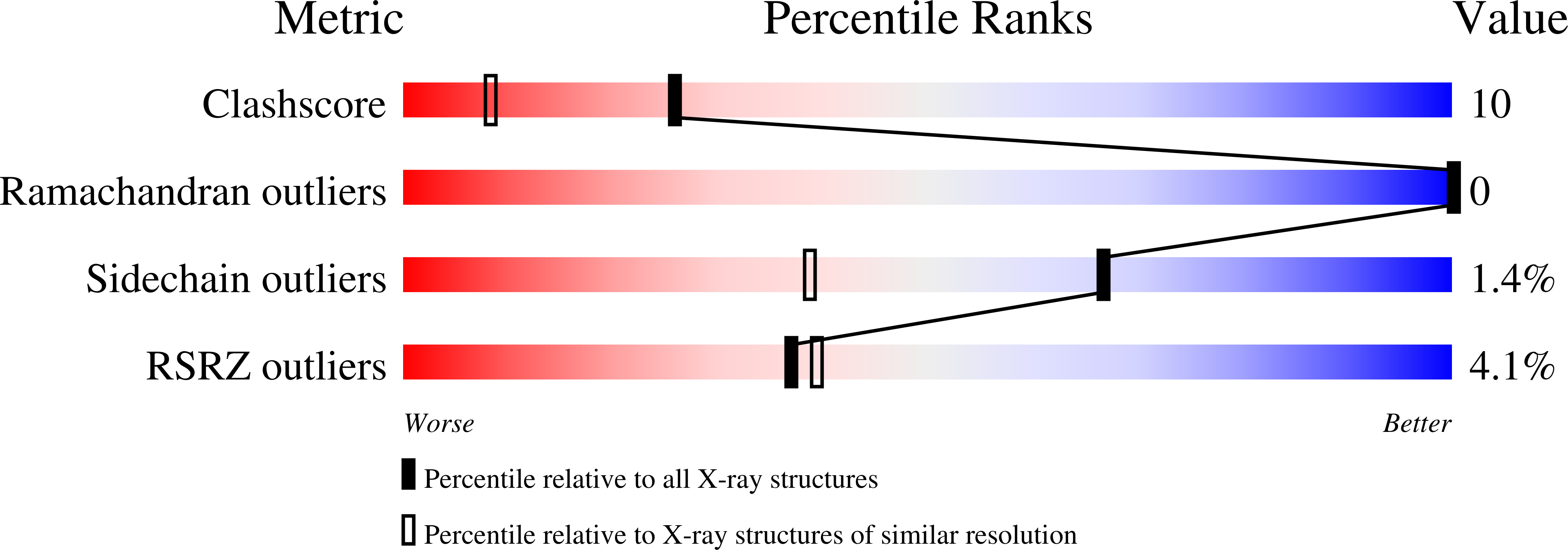
  • Method: X-RAY DIFFRACTION
  • Resolution: 1.37 Å
  • R-Value Free: 0.226 
  • R-Value Work: 0.202 
  • R-Value Observed: 0.203 

Starting Model: experimental
View more details

wwPDB Validation   3D Report Full Report


This is version 1.4 of the entry. See complete history


Literature

Structural Analysis of Haemin Demetallation by L-Chain Apoferritins

De Val, N.Declercq, J.P.Lim, C.K.Crichton, R.R.

(2012) J Inorg Biochem 112: 77

  • DOI: https://doi.org/10.1016/j.jinorgbio.2012.02.031
  • Primary Citation of Related Structures:  
    2V2I, 2V2J, 2V2L, 2V2M, 2V2N, 2V2O, 2V2P, 2V2R, 2V2S, 2W0O

  • PubMed Abstract: 

    There are extensive structural similarities between eukaryotic and prokaryotic ferritins. However, there is one essential difference between these two types of ferritins: bacterioferritins contain haem whereas eukaryotic ferritins are considered to be non-haem proteins. In vitro experiments had shown that horse spleen apoferritin or recombinant horse L chain apoferritins, when co-crystallised with haemin, undergoes demetallation of the porphyrin. In the present study a cofactor has been isolated directly from horse spleen apoferritin and from crystals of the mutant horse L chain apoferritin (E53Q, E56Q, E57Q, E60Q and R59M) which had been co-crystallised with haemin. In both cases the HPLC/ESI-MS results confirm that the cofactor is a N-ethylprotoporphyrin IX. Crystal structures of wild type L chain horse apoferritin and its three mutants co-crystallised with haemin have been determined to high resolution and in all cases a metal-free molecule derived from haemin was found in the hydrophobic pocket, close to the two-fold axis. The X-ray structure of the E53Q, E56Q, E57Q, E60Q+R59M recombinant horse L-chain apoferritin has been obtained at a higher resolution (1.16Å) than previously reported for any mammalian apoferritins. Similar evidence for a metal-free molecule derived from haemin was found in the electron density map of horse spleen apoferritin (at a resolution of 1.5Å). The out-of-plane distortion of the observed porphyrin is clearly compatible with an N-alkyl porphyrin. We conclude that L-chain ferritins are capable of binding and demetallating haemin, generating in the process N-ethylprotoporphyrin IX both in vivo and in vitro.


  • Organizational Affiliation

    Institute of Life Sciences, University of Louvain, Place Croix du Sud, 1348 Louvain-la-Neuve, Belgium. [email protected]


Macromolecules
Find similar proteins by:  (by identity cutoff)  |  3D Structure
Entity ID: 1
MoleculeChains Sequence LengthOrganismDetailsImage
FERRITIN LIGHT CHAIN174Equus caballusMutation(s): 1 
UniProt
Find proteins for P02791 (Equus caballus)
Explore P02791 
Go to UniProtKB:  P02791
Entity Groups  
Sequence Clusters30% Identity50% Identity70% Identity90% Identity95% Identity100% Identity
UniProt GroupP02791
Sequence Annotations
Expand
  • Reference Sequence
Small Molecules
Ligands 3 Unique
IDChains Name / Formula / InChI Key2D Diagram3D Interactions
CD
Query on CD

Download Ideal Coordinates CCD File 
B [auth A]
C [auth A]
D [auth A]
E [auth A]
F [auth A]
B [auth A],
C [auth A],
D [auth A],
E [auth A],
F [auth A],
G [auth A],
H [auth A],
I [auth A]
CADMIUM ION
Cd
WLZRMCYVCSSEQC-UHFFFAOYSA-N
SO4
Query on SO4

Download Ideal Coordinates CCD File 
K [auth A]SULFATE ION
O4 S
QAOWNCQODCNURD-UHFFFAOYSA-L
GOL
Query on GOL

Download Ideal Coordinates CCD File 
J [auth A],
L [auth A]
GLYCEROL
C3 H8 O3
PEDCQBHIVMGVHV-UHFFFAOYSA-N
Experimental Data & Validation

Experimental Data

  • Method: X-RAY DIFFRACTION
  • Resolution: 1.37 Å
  • R-Value Free: 0.226 
  • R-Value Work: 0.202 
  • R-Value Observed: 0.203 
  • Space Group: F 4 3 2
Unit Cell:
Length ( Å )Angle ( ˚ )
a = 181.242α = 90
b = 181.242β = 90
c = 181.242γ = 90
Software Package:
Software NamePurpose
REFMACrefinement
XDSdata reduction
XSCALEdata scaling

Structure Validation

View Full Validation Report


Entry History 

Deposition Data

Revision History  (Full details and data files)

  • Version 1.0: 2008-06-24
    Type: Initial release
  • Version 1.1: 2012-05-16
    Changes: Database references, Derived calculations, Non-polymer description, Other, Source and taxonomy, Structure summary, Version format compliance
  • Version 1.2: 2012-05-30
    Changes: Other
  • Version 1.3: 2019-07-24
    Changes: Data collection
  • Version 1.4: 2023-12-13
    Changes: Data collection, Database references, Derived calculations, Other, Refinement description