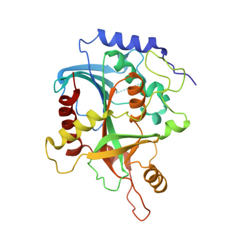
2OC9

Crystal structure of human purine nucleoside phosphorylase mutant H257G with Imm-H


Experimental Data Snapshot

  • Method: X-RAY DIFFRACTION
  • Resolution: 2.59 Å
  • R-Value Free: 0.243 
  • R-Value Work: 0.194 
  • R-Value Observed: 0.197 

Starting Model: experimental
View more details

wwPDB Validation   3D Report Full Report


Ligand Structure Quality Assessment 


This is version 1.7 of the entry. See complete history


Literature

Neighboring Group Participation in the Transition State of Human Purine Nucleoside Phosphorylase

Murkin, A.S.Birck, M.R.Rinaldo-Matthis, A.Shi, W.Taylor, E.A.Almo, S.C.Schramm, V.L.

(2007) Biochemistry 46: 5038-5049

  • DOI: https://doi.org/10.1021/bi700147b
  • Primary Citation of Related Structures:  
    2A0W, 2A0X, 2A0Y, 2OC4, 2OC9, 2ON6

  • PubMed Abstract: 

    The X-ray crystal structures of human purine nucleoside phosphorylase (PNP) with bound inosine or transition-state analogues show His257 within hydrogen bonding distance of the 5'-hydroxyl. The mutants His257Phe, His257Gly, and His257Asp exhibited greatly decreased affinity for Immucillin-H (ImmH), binding this mimic of an early transition state as much as 370-fold (Km/Ki) less tightly than native PNP. In contrast, these mutants bound DADMe-ImmH, a mimic of a late transition state, nearly as well as the native enzyme. These results indicate that His257 serves an important role in the early stages of transition-state formation. Whereas mutation of His257 resulted in little variation in the PNP x DADMe-ImmH x SO4 structures, His257Phe x ImmH x PO4 showed distortion at the 5'-hydroxyl, indicating the importance of H-bonding in positioning this group during progression to the transition state. Binding isotope effect (BIE) and kinetic isotope effect (KIE) studies of the remote 5'-(3)H for the arsenolysis of inosine with native PNP revealed a BIE of 1.5% and an unexpectedly large intrinsic KIE of 4.6%. This result is interpreted as a moderate electronic distortion toward the transition state in the Michaelis complex with continued development of a similar distortion at the transition state. The mutants His257Phe, His257Gly, and His257Asp altered the 5'-(3)H intrinsic KIE to -3, -14, and 7%, respectively, while the BIEs contributed 2, 2, and -2%, respectively. These surprising results establish that forces in the Michaelis complex, reported by the BIEs, can be reversed or enhanced at the transition state.


  • Organizational Affiliation

    Department of Biochemistry, Albert Einstein College of Medicine, 1300 Morris Park Avenue, Bronx, New York 10461, USA.


Macromolecules
Find similar proteins by:  (by identity cutoff)  |  3D Structure
Entity ID: 1
MoleculeChains Sequence LengthOrganismDetailsImage
Purine nucleoside phosphorylase289Homo sapiensMutation(s): 2 
Gene Names: NPPNP
EC: 2.4.2.1
UniProt & NIH Common Fund Data Resources
Find proteins for P00491 (Homo sapiens)
Explore P00491 
Go to UniProtKB:  P00491
PHAROS:  P00491
GTEx:  ENSG00000198805 
Entity Groups  
Sequence Clusters30% Identity50% Identity70% Identity90% Identity95% Identity100% Identity
UniProt GroupP00491
Sequence Annotations
Expand
  • Reference Sequence
Small Molecules
Ligands 2 Unique
IDChains Name / Formula / InChI Key2D Diagram3D Interactions
IMH
Query on IMH

Download Ideal Coordinates CCD File 
C [auth A]1,4-DIDEOXY-4-AZA-1-(S)-(9-DEAZAHYPOXANTHIN-9-YL)-D-RIBITOL
C11 H14 N4 O4
IWKXDMQDITUYRK-KUBHLMPHSA-N
PO4
Query on PO4

Download Ideal Coordinates CCD File 
B [auth A]PHOSPHATE ION
O4 P
NBIIXXVUZAFLBC-UHFFFAOYSA-K
Binding Affinity Annotations 
IDSourceBinding Affinity
IMH BindingDB:  2OC9 Ki: min: 0.06, max: 72 (nM) from 6 assay(s)
Kd: min: 0.02, max: 0.06 (nM) from 3 assay(s)
IC50: min: 0.06, max: 100 (nM) from 4 assay(s)
ΔH: -3.50e+1 (kJ/mol) from 1 assay(s)
ΔG: -4.56e+1 (kJ/mol) from 1 assay(s)
PDBBind:  2OC9 Ki: 11 (nM) from 1 assay(s)
Experimental Data & Validation

Experimental Data

  • Method: X-RAY DIFFRACTION
  • Resolution: 2.59 Å
  • R-Value Free: 0.243 
  • R-Value Work: 0.194 
  • R-Value Observed: 0.197 
  • Space Group: H 3 2
Unit Cell:
Length ( Å )Angle ( ˚ )
a = 142.17α = 90
b = 142.17β = 90
c = 168.674γ = 120
Software Package:
Software NamePurpose
DENZOdata reduction
SCALEPACKdata scaling
MOLREPphasing
REFMACrefinement
PDB_EXTRACTdata extraction
CBASSdata collection
HKL-2000data reduction

Structure Validation

View Full Validation Report


Ligand Structure Quality Assessment 


Entry History 

Deposition Data

Revision History  (Full details and data files)

  • Version 1.0: 2007-05-22
    Type: Initial release
  • Version 1.1: 2008-05-01
    Changes: Version format compliance
  • Version 1.2: 2011-07-13
    Changes: Derived calculations, Version format compliance
  • Version 1.3: 2017-02-01
    Changes: Structure summary
  • Version 1.4: 2017-10-18
    Changes: Refinement description
  • Version 1.5: 2021-10-20
    Changes: Database references, Derived calculations
  • Version 1.6: 2022-10-05
    Changes: Structure summary
  • Version 1.7: 2023-09-20
    Changes: Data collection, Refinement description