2LBD

LIGAND-BINDING DOMAIN OF THE HUMAN RETINOIC ACID RECEPTOR GAMMA BOUND TO ALL-TRANS RETINOIC ACID


Experimental Data Snapshot

  • Method: X-RAY DIFFRACTION
  • Resolution: 2.06 Å
  • R-Value Free: 
    0.313 (Depositor), 0.300 (DCC) 
  • R-Value Work: 
    0.210 (Depositor), 0.210 (DCC) 
  • R-Value Observed: 
    0.210 (Depositor) 

wwPDB Validation   3D Report Full Report


Ligand Structure Quality Assessment 

Created with Raphaël 2.3.0Worse 01 BetterLigand structure goodness of fit to experimental dataBest fitted REAClick on this verticalbar to view details

This is version 1.3 of the entry. See complete history


Literature

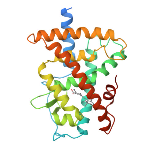
Crystal structure of the RAR-gamma ligand-binding domain bound to all-trans retinoic acid.

Renaud, J.P.Rochel, N.Ruff, M.Vivat, V.Chambon, P.Gronemeyer, H.Moras, D.

(1995) Nature 378: 681-689

  • DOI: https://doi.org/10.1038/378681a0
  • Primary Citation of Related Structures:  
    2LBD

  • PubMed Abstract: 

    The 2.0-A crystal structure of the ligand-binding domain (LBD) of the human retinoic acid receptor (RAR)-gamma bound to all-trans retinoic acid reveals the ligand-binding interactions and suggests an electrostatic guidance mechanism. The overall fold is similar to that of the human RXR-alpha apo-LBD, except for the carboxy-terminal part which folds back towards the LBD core, contributing to the hydrophobic ligand pocket and 'sealing' its entry site. We propose a 'mouse trap' mechanism whereby a ligand-induced conformational transition repositions the amphipathic alpha-helix of the AF-2 activating domain and forms a transcriptionally active receptor.


  • Organizational Affiliation

    Institut de Génétique et de Biologie Moléculaire et Cellulaire, CNRS/INSERM/ULP/Collège de France, Strasbourg, France.


Macromolecules
Find similar proteins by:  (by identity cutoff)  |  3D Structure
Entity ID: 1
MoleculeChains Sequence LengthOrganismDetailsImage
RETINOIC ACID RECEPTOR GAMMA267Homo sapiensMutation(s): 0 
Gene Names: HUMAN RAR GAMMA A CDNA (NUCLEOTIDES 946 - 1683)
UniProt & NIH Common Fund Data Resources
Find proteins for P13631 (Homo sapiens)
Explore P13631 
Go to UniProtKB:  P13631
PHAROS:  P13631
GTEx:  ENSG00000172819 
Entity Groups  
Sequence Clusters30% Identity50% Identity70% Identity90% Identity95% Identity100% Identity
UniProt GroupP13631
Sequence Annotations
Expand
  • Reference Sequence
Small Molecules
Ligands 1 Unique
IDChains Name / Formula / InChI Key2D Diagram3D Interactions
REA
Query on REA

Download Ideal Coordinates CCD File 
B [auth A]RETINOIC ACID
C20 H28 O2
SHGAZHPCJJPHSC-YCNIQYBTSA-N
Binding Affinity Annotations 
IDSourceBinding Affinity
REA BindingDB:  2LBD IC50: min: 1000, max: 1000 (nM) from 2 assay(s)
Experimental Data & Validation

Experimental Data

  • Method: X-RAY DIFFRACTION
  • Resolution: 2.06 Å
  • R-Value Free:  0.313 (Depositor), 0.300 (DCC) 
  • R-Value Work:  0.210 (Depositor), 0.210 (DCC) 
  • R-Value Observed: 0.210 (Depositor) 
Space Group: P 41 21 2
Unit Cell:
Length ( Å )Angle ( ˚ )
a = 60.6α = 90
b = 60.6β = 90
c = 155.3γ = 90
Software Package:
Software NamePurpose
X-PLORmodel building
X-PLORrefinement
MARXDSdata reduction
MARSCALEdata scaling
X-PLORphasing

Structure Validation

View Full Validation Report


Ligand Structure Quality Assessment 

Created with Raphaël 2.3.0Worse 01 BetterLigand structure goodness of fit to experimental dataBest fitted REAClick on this verticalbar to view details

Entry History 

Deposition Data

Revision History  (Full details and data files)

  • Version 1.0: 1997-11-12
    Type: Initial release
  • Version 1.1: 2008-03-24
    Changes: Version format compliance
  • Version 1.2: 2011-07-13
    Changes: Version format compliance
  • Version 1.3: 2024-02-21
    Changes: Data collection, Database references, Derived calculations, Other