2F9N

Crystal Structure of the Recombinant Human Alpha I Tryptase Mutant K192Q/D216G in Complex with Leupeptin


Experimental Data Snapshot

  • Method: X-RAY DIFFRACTION
  • Resolution: 1.60 Å
  • R-Value Free: 0.236 
  • R-Value Work: 0.190 
  • R-Value Observed: 0.190 

Starting Model: experimental
View more details

wwPDB Validation   3D Report Full Report


Ligand Structure Quality Assessment 

Created with Raphaël 2.3.0Worse 01 BetterLigand structure goodness of fit to experimental dataBest fitted NAGClick on this verticalbar to view details

This is version 2.3 of the entry. See complete history


Literature

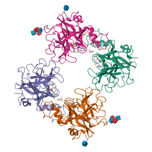
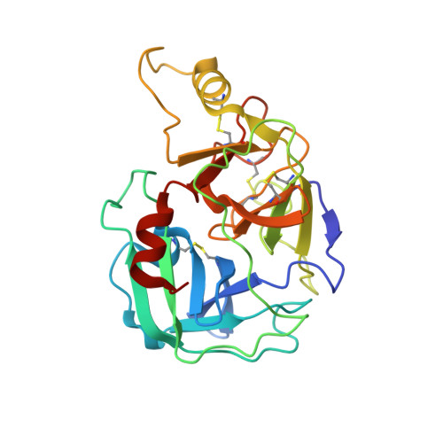
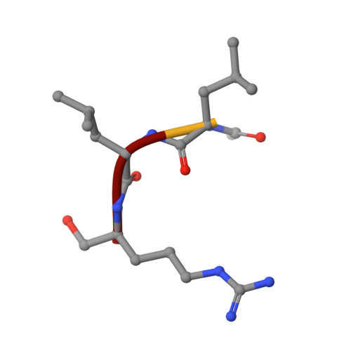
X-ray Structures of Free and Leupeptin-complexed Human alpha I-Tryptase Mutants: Indication for an alpha to beta-Tryptase Transition

Rohr, K.B.Selwood, T.Marquardt, U.Huber, R.Schechter, N.M.Bode, W.Than, M.E.

(2005) J Mol Biol 357: 195-209

  • DOI: https://doi.org/10.1016/j.jmb.2005.12.037
  • Primary Citation of Related Structures:  
    2F9N, 2F9O, 2F9P

  • PubMed Abstract: 

    Tryptases alpha and beta are trypsin-like serine proteinases expressed in large amounts by mast cells. Beta-tryptase is a tetramer that has enzymatic activity, but requires heparin binding to maintain functional and structural stability, whereas alpha-tryptase has little, if any, enzymatic activity but is a stable tetramer in the absence of heparin. As shown previously, these differences can be mainly attributed to the different conformations of the 214-220 segment. Interestingly, the replacement of Asp216 by Gly, which is present in beta-tryptase, results in enzymatically active but less stable alpha-tryptase mutants. We have solved the crystal structures of both the single (D216G) and the double (K192Q/D216G) mutant forms of recombinant human alphaI-tryptase in complex with the peptide inhibitor leupeptin, as well as the structure of the non-inhibited single mutant. The inhibited mutants exhibited an open functional substrate binding site, while in the absence of an inhibitor, the open (beta-tryptase-like) and the closed (alpha-tryptase-like) conformations were present simultaneously. This shows that both forms are in a two-state equilibrium, which is influenced by the residues in the vicinity of the active site and by inhibitor/substrate binding. Novel insights regarding the observed stability differences as well as a potential proteolytic activity of wild-type alpha-tryptase, which may possess a cryptic active site, are discussed.


  • Organizational Affiliation

    Max-Planck-Institut für Biochemie, Abteilung Strukturforschung, Am Klopferspitz 18, 82152 Martinsried, Germany.


Macromolecules
Find similar proteins by:  (by identity cutoff)  |  3D Structure
Entity ID: 1
MoleculeChains Sequence LengthOrganismDetailsImage
alpha I tryptase
A, B, C, D
245Homo sapiensMutation(s): 5 
Gene Names: TPS1TPS2TPSAB1TPSB1
EC: 3.4.21.59
UniProt & NIH Common Fund Data Resources
Find proteins for Q15661 (Homo sapiens)
Explore Q15661 
Go to UniProtKB:  Q15661
PHAROS:  Q15661
GTEx:  ENSG00000172236 
Entity Groups  
Sequence Clusters30% Identity50% Identity70% Identity90% Identity95% Identity100% Identity
UniProt GroupQ15661
Glycosylation
Glycosylation Sites: 2Go to GlyGen: Q15661-1
Sequence Annotations
Expand
  • Reference Sequence

Find similar proteins by:  Sequence   |   3D Structure  

Entity ID: 2
MoleculeChains Sequence LengthOrganismDetailsImage
Leupeptin
E, F, G, H
4Streptomyces roseusMutation(s): 0 
Sequence Annotations
Expand
  • Reference Sequence
Oligosaccharides

Help

Entity ID: 3
MoleculeChains Length2D Diagram Glycosylation3D Interactions
alpha-L-fucopyranose-(1-3)-[2-acetamido-2-deoxy-beta-D-glucopyranose-(1-4)][alpha-L-fucopyranose-(1-6)]2-acetamido-2-deoxy-beta-D-glucopyranose
I, J
4N-Glycosylation
Glycosylation Resources
GlyTouCan:  G63564LA
GlyCosmos:  G63564LA
Experimental Data & Validation

Experimental Data

  • Method: X-RAY DIFFRACTION
  • Resolution: 1.60 Å
  • R-Value Free: 0.236 
  • R-Value Work: 0.190 
  • R-Value Observed: 0.190 
  • Space Group: P 21 21 21
Unit Cell:
Length ( Å )Angle ( ˚ )
a = 83.31α = 90
b = 88.57β = 90
c = 163.37γ = 90
Software Package:
Software NamePurpose
DENZOdata reduction
SCALEPACKdata scaling
AMoREphasing
CNSrefinement

Structure Validation

View Full Validation Report


Ligand Structure Quality Assessment 

Created with Raphaël 2.3.0Worse 01 BetterLigand structure goodness of fit to experimental dataBest fitted NAGClick on this verticalbar to view details

Entry History 

Deposition Data

Revision History  (Full details and data files)

  • Version 1.0: 2006-01-31
    Type: Initial release
  • Version 1.1: 2008-05-01
    Changes: Version format compliance
  • Version 1.2: 2011-07-13
    Changes: Atomic model, Database references, Derived calculations, Non-polymer description, Structure summary, Version format compliance
  • Version 1.3: 2012-12-12
    Changes: Other
  • Version 2.0: 2020-07-29
    Type: Remediation
    Reason: Carbohydrate remediation
    Changes: Advisory, Atomic model, Data collection, Database references, Derived calculations, Structure summary
  • Version 2.1: 2021-10-20
    Changes: Advisory, Database references, Structure summary
  • Version 2.2: 2023-08-30
    Changes: Data collection, Refinement description
  • Version 2.3: 2024-10-30
    Changes: Structure summary