


Crystal Structure of T.th.HB8 Branched-Chain Amino Acid Aminotransferase Complexed with 4-Methylvaleric Acid
Goto, M.To be published.
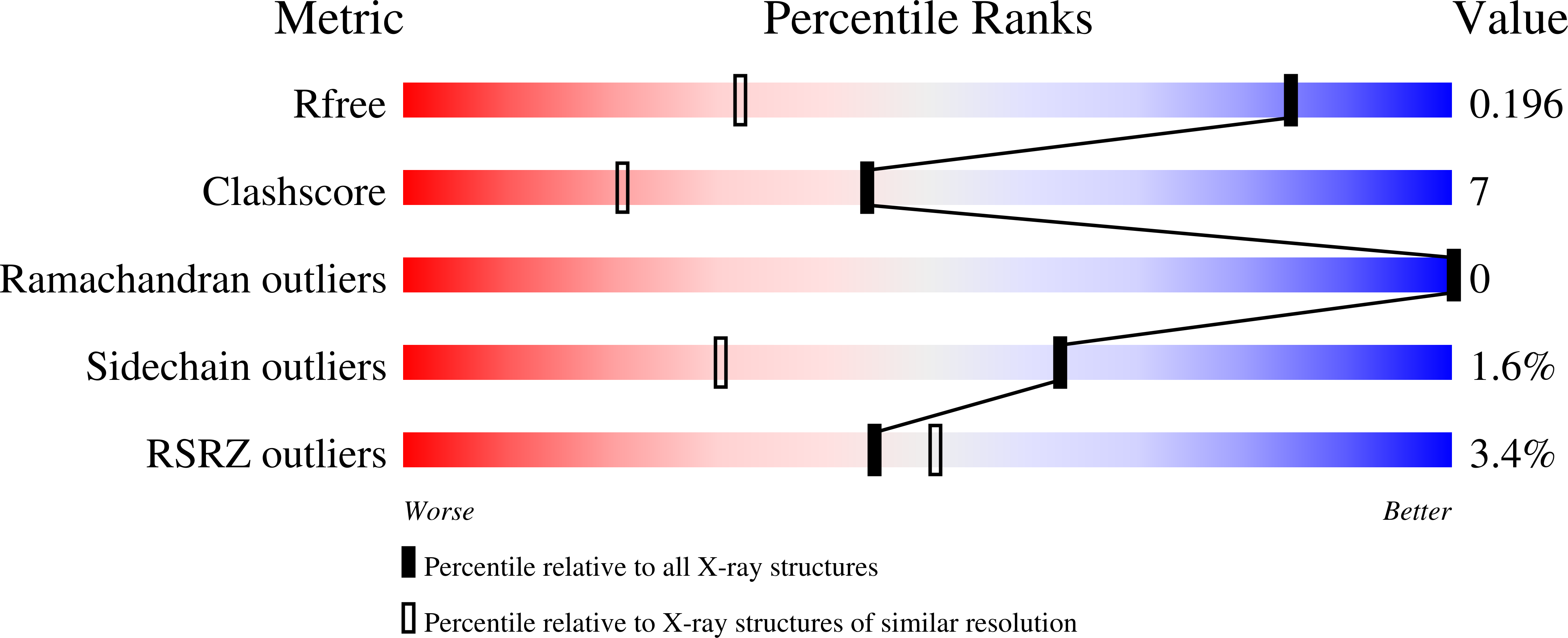
Experimental Data Snapshot
Starting Model: experimental
View more details
Entity ID: 1 | |||||
|---|---|---|---|---|---|
| Molecule | Chains | Sequence Length | Organism | Details | Image |
| Branched-chain amino acid aminotransferase | 308 | Thermus thermophilus HB8 | Mutation(s): 0 EC: 2.6.1.42 | 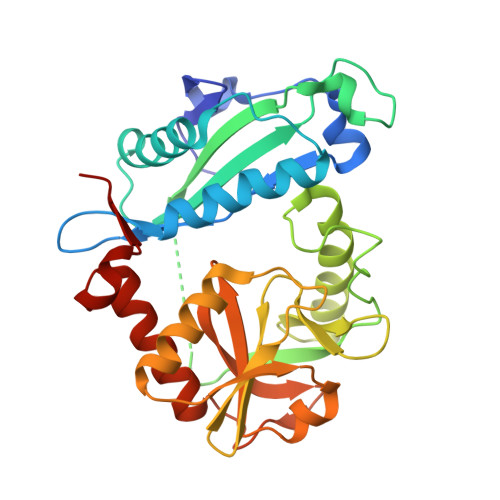 | |
UniProt | |||||
Find proteins for Q5SM19 (Thermus thermophilus (strain ATCC 27634 / DSM 579 / HB8)) Explore Q5SM19 Go to UniProtKB: Q5SM19 | |||||
Entity Groups | |||||
| Sequence Clusters | 30% Identity50% Identity70% Identity90% Identity95% Identity100% Identity | ||||
| UniProt Group | Q5SM19 | ||||
Sequence AnnotationsExpand | |||||
| |||||
| Ligands 3 Unique | |||||
|---|---|---|---|---|---|
| ID | Chains | Name / Formula / InChI Key | 2D Diagram | 3D Interactions | |
| PLP Query on PLP | D [auth A], H [auth B], L [auth C] | PYRIDOXAL-5'-PHOSPHATE C8 H10 N O6 P NGVDGCNFYWLIFO-UHFFFAOYSA-N |  | ||
| MPD Query on MPD | F [auth A], G [auth A], J [auth B], K [auth B], N [auth C] | (4S)-2-METHYL-2,4-PENTANEDIOL C6 H14 O2 SVTBMSDMJJWYQN-YFKPBYRVSA-N |  | ||
| 4MV Query on 4MV | E [auth A], I [auth B], M [auth C] | 4-METHYL VALERIC ACID C6 H12 O2 FGKJLKRYENPLQH-UHFFFAOYSA-N |  | ||
| Length ( Å ) | Angle ( ˚ ) |
|---|---|
| a = 143.652 | α = 90 |
| b = 143.652 | β = 90 |
| c = 116.54 | γ = 90 |
| Software Name | Purpose |
|---|---|
| CNS | refinement |
| MAR345 | data collection |
| MOSFLM | data reduction |
| SCALA | data scaling |
| AMoRE | phasing |
RCSB PDB is hosted by

RCSB PDB is a member of the


